. 2016 Jun 21:326:84-94.
doi: 10.1016/j.neuroscience.2016.03.054. Epub 2016 Apr 4.

Unilateral microinjection of acrolein into thoracic spinal cord produces acute and chronic injury and functional deficits

Affiliations

Unilateral microinjection of acrolein into thoracic spinal cord produces acute and chronic injury and functional deficits

Alexander Gianaris et al. Neuroscience. .

Abstract

Although lipid peroxidation has long been associated with spinal cord injury (SCI), the specific role of lipid peroxidation-derived byproducts such as acrolein in mediating damage remains to be fully understood. Acrolein, an α-β unsaturated aldehyde, is highly reactive with proteins, DNA, and phospholipids and is considered as a second toxic messenger that disseminates and augments initial free radical events. Previously, we showed that acrolein increased following traumatic SCI and injection of acrolein induced tissue damage. Here, we demonstrate that microinjection of acrolein into the thoracic spinal cord of adult rats resulted in dose-dependent tissue damage and functional deficits. At 24h (acute) after the microinjection, tissue damage, motoneuron loss, and spinal cord swelling were observed on sections stained with Cresyl Violet. Luxol fast blue staining further showed that acrolein injection resulted in dose-dependent demyelination. At 8weeks (chronic) after the microinjection, cord shrinkage, astrocyte activation, and macrophage infiltration were observed along with tissue damage, neuron loss, and demyelination. These pathological changes resulted in behavioral impairments as measured by both the Basso, Beattie, and Bresnahan (BBB) locomotor rating scale and grid walking analysis. Electron microscopy further demonstrated that acrolein induced axonal degeneration, demyelination, and macrophage infiltration. These results, combined with our previous reports, strongly suggest that acrolein may play a critical causal role in the pathogenesis of SCI and that targeting acrolein could be an attractive strategy for repair after SCI.

Keywords: acrolein; aldehyde; lipid peroxidation; oxidative stress; spinal cord injury.


Figures

Figure 1
Figure 1
Diagram of microinjection and experimental timeline. Saline or acrolein was injected into the rat spinal cord at a depth of 1.3 mm and a distance of 0.6 mm lateral from the midline at T10. Basso, Beattie, and Bresnahan (BBB) locomotor rating scale and grid walking tests were performed prior to the injection procedure, 24 h after injection, and then once a week for 7 wk. Animals were sacrificed at 24 h (acute observation) and 8 wk (chronic observation), respectively, after the injection.
Figure 2
Figure 2
Basso, Beattie, and Bresnahan (BBB) locomotor rating scale score over 7 wk after injections of acrolein into the normal spinal cord of adult rats. The BBB locomotion rating scale showed that BBB scores decreased in response to increased doses of acrolein (**: p<0.01, vs saline; #: p<0.05, ##: p<0.01, vs 0.1μmol acrolein).
Figure 3
Figure 3
Foot drops via grid walking test over 7 wk after injections of acrolein into the normal adult rat spinal cord on the right side at T10. (A) Significant increases of foot drops on the side ipsilateral to the acrolein injection were found in both doses (0.1 and 1.0 μmol) at 24 h after the injection. At most time points (except for the 4th wk), injection of high dose acrolein (1.0 μmol) resulted in significantly greater foot drops on the injury side than the low dose (0.1 μmol) or saline control (*: p<0.05,**: p<0.01, vs saline; #: p<0.05, vs 0.1μmol acrolein). (B) The foot drops on the side contralateral to the injection showed no difference amongst groups.
Figure 4
Figure 4
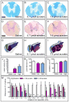
Acrolein induced graded tissue damage at 24 h after injection. (A, D) Luxol fast blue (A) and Cresyl violet-eosin (D) stainings show no tissue damage or demyelination in a saline-injected spinal cord. (B, C, E, F) Low (0.1 μmol, B, E) and high dose (1.0 μmol, C, F) acrolein injections induced a confined lesion and demyelination in the ventral and ventrolateral grey and white matter. Bars: A–F, 500 μm. (G–I) Representative three-dimensional reconstruction of a spinal cord segment from each group illustrates rostrocaudal extension of the lesion (red). (J–L) Bar graphs show acrolein injection induced percent changes in lesion volume (J), demyelination (K), and cord swelling (L) among the three groups (Low, 0.1 μmol acrolein; High, 1.0 μmol acrolein; *: p<0.05,**: p<0.01, vs saline). (M) Acrolein injections into the right spinal cord cause significant dose-related motoneuron loss (*: p<0.05, **: p<0.01, vs saline; ##: p<0.01, vs 0.1μmol acrolein).
Figure 5
Figure 5
Injections of acrolein into the normal spinal cord resulted in tissue damage in a dose-dependent manner at 8 wk. (A, D) Luxol fast blue (A) and Cresyl violet-eosin (D) stainings showed no tissue damage or demyelination in a saline-injected spinal cord. (B, C, E, F) Low (0.1 μmol, B, E) and high dose (1.0 μmol, C, F) acrolein injections induced a confined lesion and demyelination in the ventral and ventrolateral grey and white matter. Bars: A–F, 500 μm. (G–I) Representative three-dimensional reconstruction of a spinal cord segment from each group illustrates rostrocaudal extension of the lesion (red). (J–L) Bar graphs show acrolein injection induced percent changes in lesion volume (J), demyelination (K), and cord swelling (L) among the three groups (Low, 0.1 μmol acrolein; High, 1.0 μmol acrolein; *: p<0.05,**: p<0.01, vs saline; ##: p<0.01, vs 0.1 μmole acrolein). (M) Acrolein injections into the right spinal cord caused significant dose-related motoneuron loss (*: p<0.05, **: p<0.01, vs saline; #: p<0.05, ##: p<0.01, vs 0.1 μmole acrolein).
Figure 6
Figure 6
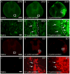
Immunofluorescence staining at 8th wk after acrolein injection. (A–F) Increased GFAP expression at the lesion border shows acrolein-induced reactive gliosis in both low (0.1 μmol) and high (1.0 μmol) doses of acrolein injection groups. (D–F) High magnification of boxed areas in A-C shows normal (D) and reactive (E, F, arrows) astrocytes. Within the lesion epicenter, non-specifically labeled, morphologically characteristic of macrophages were also found (F, arrowheads). (G–I) Representative photomicrographs show increased ED-1 expression, a macrophage marker, within the lesion site of low and high doses of acrolein injections. (J–L) High magnification of boxed areas of G–I showed the lack of (J) or presence of (K, L, arrows) macrophages stained with ED-1 after saline (J) or acrolein (K, L) injections. The invaded macrophages were confined mainly within the lesion site. Bars: A–C and G–I, 500 μm; D–F and J–L, 40 μm.
Figure 7
Figure 7
Electron micrographs after acrolein injection. Representative photographs of electron microscopic images show normal appearance of axons and myelin at 24 h and 8 wk after saline injection (A, D). Low and high dose of acrolein injection, 0.1 μmol and 1.0 μmol, respectively, induced axon-myelin pathology including large axon with thin myelin (B, F, double arrows), axon degeneration (C, arrows), axon and myelin degeneration (E, arrows), and macrophage engulfment of degenerated cell debris (F, arrow head) at 24 h (A–C) and 8 wk (D–F) after acrolein injection. Bar = 6 μm.

References

    1. Spinal cord injury facts and figures at a glance. J Spinal Cord Med. 2012;35:480–481. - PMC - PubMed
    1. Basso DM, Beattie MS, Bresnahan JC. A sensitive and reliable locomotor rating scale for open field testing in rats. J Neurotrauma. 1995;12:1–21. - PubMed
    1. Basso DM, Beattie MS, Bresnahan JC. Graded histological and locomotor outcomes after spinal cord contusion using the NYU weight-drop device versus transection. Exp Neurol. 1996;139:244–256. - PubMed
    1. Behrmann DL, Bresnahan JC, Beattie MS, Shah BR. Spinal cord injury produced by consistent mechanical displacement of the cord in rats: behavioral and histologic analysis. J Neurotrauma. 1992;9:197–217. - PubMed
    1. Blight AR. Delayed demyelination and macrophage invasion: a candidate for secondary cell damage in spinal cord injury. Cent Nerv Syst Trauma. 1985;2:299–315. - PubMed

Publication types