

Somatic Activating Mutations in GNAQ and GNA11 Are Associated with Congenital Hemangioma
- PMID: 27058448
- PMCID: PMC4833432
- DOI: 10.1016/j.ajhg.2016.03.009
Somatic Activating Mutations in GNAQ and GNA11 Are Associated with Congenital Hemangioma
Erratum in
-
Somatic Activating Mutations in GNAQ and GNA11 Are Associated with Congenital Hemangioma.Am J Hum Genet. 2016 Jun 2;98(6):1271. doi: 10.1016/j.ajhg.2016.05.010. Epub 2016 Jun 2. Am J Hum Genet. 2016. PMID: 27259057 Free PMC article. No abstract available.
Abstract
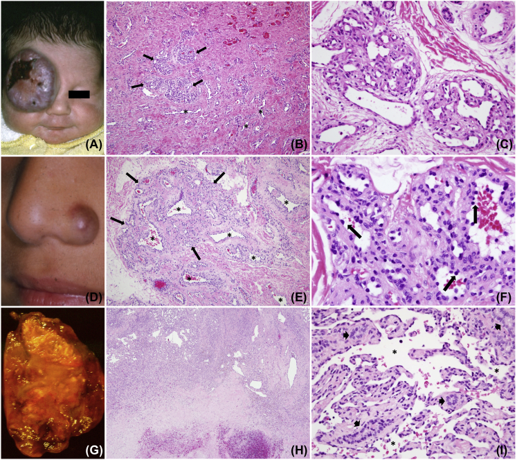
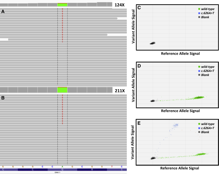
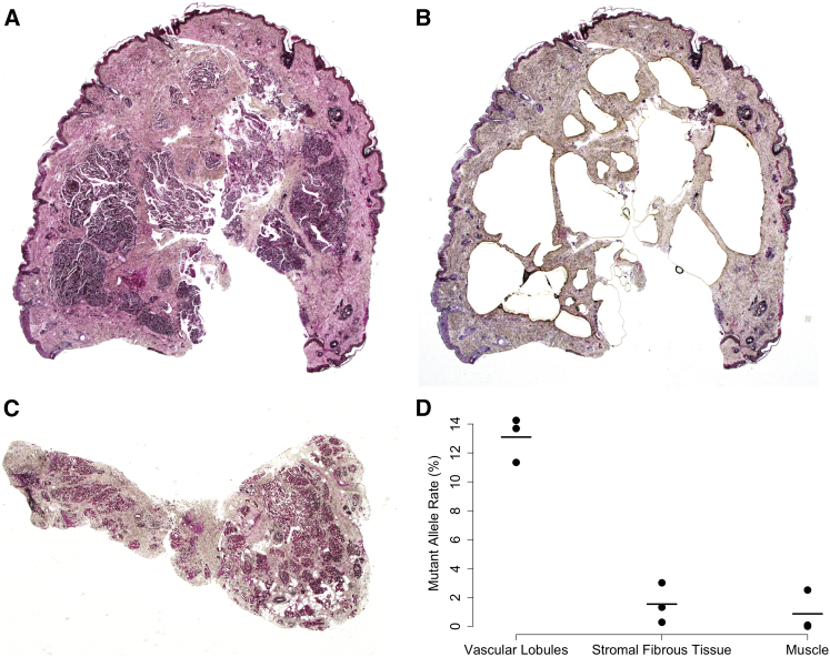
Congenital hemangioma is a rare vascular tumor that forms in utero. Postnatally, the tumor either involutes quickly (i.e., rapidly involuting congenital hemangioma [RICH]) or partially regresses and stabilizes (i.e., non-involuting congenital hemangioma [NICH]). We hypothesized that congenital hemangiomas arise due to somatic mutation and performed massively parallel mRNA sequencing on affected tissue from eight participants. We identified mutually exclusive, mosaic missense mutations that alter glutamine at amino acid 209 (Glu209) in GNAQ or GNA11 in all tested samples, at variant allele frequencies (VAF) ranging from 3% to 33%. We verified the presence of the mutations in genomic DNA using a combination of molecular inversion probe sequencing (MIP-seq) and digital droplet PCR (ddPCR). The Glu209 GNAQ and GNA11 missense variants we identified are common in uveal melanoma and have been shown to constitutively activate MAPK and/or YAP signaling. When we screened additional archival formalin-fixed paraffin-embedded (FFPE) congenital cutaneous and hepatic hemangiomas, 4/8 had GNAQ or GNA11 Glu209 variants. The same GNAQ or GNA11 mutation is found in both NICH and RICH, so other factors must account for these tumors' different postnatal behaviors.
Copyright © 2016 The American Society of Human Genetics. Published by Elsevier Inc. All rights reserved.
Figures
References
-
- North P.E., Waner M., Mizeracki A., Mihm M.C., Jr. GLUT1: a newly discovered immunohistochemical marker for juvenile hemangiomas. Hum. Pathol. 2000;31:11–22. - PubMed
-
- Berenguer B., Mulliken J.B., Enjolras O., Boon L.M., Wassef M., Josset P., Burrows P.E., Perez-Atayde A.R., Kozakewich H.P. Rapidly involuting congenital hemangioma: clinical and histopathologic features. Pediatr. Dev. Pathol. 2003;6:495–510. - PubMed
-
- Boon L.M., Enjolras O., Mulliken J.B. Congenital hemangioma: evidence of accelerated involution. J. Pediatr. 1996;128:329–335. - PubMed
-
- Enjolras O., Mulliken J.B., Boon L.M., Wassef M., Kozakewich H.P., Burrows P.E. Noninvoluting congenital hemangioma: a rare cutaneous vascular anomaly. Plast. Reconstr. Surg. 2001;107:1647–1654. - PubMed
-
- Fishman S.J., Burrows P.E. Treatment of visceral vascular tumors. In: Mulliken J.B., Burrows P.E., Fishman S.J., editors. Mulliken and Young’s Vascular Anomalies: Hemangiomas and Malformations. Oxford University Press; 2013. pp. 242–245.
Publication types
MeSH terms
Substances
Grants and funding
LinkOut - more resources
Full Text Sources
Other Literature Sources
Medical
Molecular Biology Databases
