. 2016 Oct;68(10):2492-502.
doi: 10.1002/art.39706.

Autoantibody-Positive Healthy Individuals Display Unique Immune Profiles That May Regulate Autoimmunity

Affiliations

Autoantibody-Positive Healthy Individuals Display Unique Immune Profiles That May Regulate Autoimmunity

Samantha Slight-Webb et al. Arthritis Rheumatol. 2016 Oct.

Abstract

Objective: Antinuclear antibodies (ANAs) are detected in ∼18% of females, yet autoimmune disease develops in only 5-8%. Immunologic differences between ANA-positive healthy individuals and patients with systemic lupus erythematosus (SLE) may elucidate the regulatory mechanisms by which ANA-positive individuals avoid transition to clinical autoimmune disease.

Methods: Healthy individuals (n = 790) were screened for autoantibodies specific for 11 antigens associated with lupus, systemic sclerosis, and Sjögren's syndrome. From this screening, 31 European American ANA-positive healthy individuals were selected and demographically matched to ANA-negative controls and SLE patients. Serum cytokine profiles, leukocyte subset frequency, and reactivity were analyzed by multiplex assays, immunophenotyping, and phosphospecific flow cytometry.

Results: Of 790 individuals screened, 57 (7%) were ANA-positive. The majority of proinflammatory cytokines, including interferon-γ (IFNγ), tumor necrosis factor, interleukin-17 (IL-17), and granulocyte colony-stimulating factor, exhibited a stepwise increase in serum levels from ANA-negative controls to ANA-positive healthy individuals to SLE patients (P < 0.0001). IFNα, IFNβ, IL-12p40, and stem cell factor/c-Kit ligand were increased in SLE patients only (P < 0.05). B lymphocyte stimulator (BlyS) was elevated in SLE patients but decreased in ANA-positive individuals (P < 0.001). Further, IL-1 receptor antagonist (IL-1Ra) was down-regulated in SLE patients only (P < 0.0001). ANA-positive individuals had increased frequencies of monocytes, memory B cells, and plasmablasts and increased levels of pSTAT-1 and pSTAT-3 following IFNα stimulation compared with ANA-negative controls (P < 0.05).

Conclusion: ANA-positive healthy individuals exhibit dysregulation in multiple immune pathways yet differ from SLE patients by the absence of elevated IFNs, BLyS, IL-12p40, and stem cell factor/c-Kit ligand. Further, severely decreased levels of IL-1Ra in SLE patients compared with ANA-positive individuals may contribute to disease development. These results highlight the importance of IFN-related pathways and regulatory elements in SLE pathogenesis.


Conflict of interest statement

No conflicts of interest to report

Figures
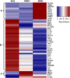
Figure 1
Figure 1
Antinuclear antibody−positive (ANA+) healthy individuals have a distinct serum soluble mediator profile. Columns represent grouped study participants: patients with systemic lupus erythematosus (SLE), ANA‐positive healthy individuals, and ANA‐negative (ANA−) healthy individuals. Rows represent individual cytokines measured by a multiplex Luminex bead‐based assay or by enzyme‐linked immunosorbent assay (B lymphocyte stimulator [BlyS] and interleukin‐1 receptor antagonist [IL‐1Ra]). In generating the heatmap, each cytokine row was normalized to have a mean of 0 and a variance of 1. Normalized cytokine values are displayed on a color scale ranging from blue (levels below the mean) through white (levels equal to the mean) to red (levels greater than the mean). Cluster A indicates soluble mediators that are down‐regulated in SLE and/or ANA‐positive healthy individuals, cluster B shows soluble mediators that are elevated in SLE and/or ANA‐positive healthy individuals, and cluster C shows soluble mediators that are up‐regulated in ANA‐positive healthy individuals only. PAI‐1 = plasminogen activator inhibitor 1; sFasL = soluble FasL; TNFβ = tumor necrosis factor β; M‐CSF = macrophage colony‐stimulating factor; VCAM‐1 = vascular cell adhesion molecule 1; ICAM‐1 = intercellular adhesion molecule 1; IFNβ = interferon‐β; TGFα = transforming growth factor α; MCP‐3 = monocyte chemotactic protein 3; Kit‐LG = Kit ligand; G‐CSF = granulocyte‐CSF; FGFβ = fibroblast growth factor β; IP‐10 = IFNγ‐inducible 10‐kd protein; NGF = nerve growth factor; PDGFBB = platelet‐derived growth factor BB; LIF = leukemia inhibitory factor; VEGF = vascular endothelial growth factor; MIP‐1β = macrophage inflammatory protein 1β.
Figure 2
Figure 2
ANA‐positive healthy individuals have higher levels of certain proinflammatory soluble mediators seen in SLE patients and elevated levels of the regulatory cytokine IL‐1Ra. Proinflammatory soluble mediators were measured by multiplex assay or enzyme‐linked immunosorbent assay and included TNF (A), IFNγ (B), IL‐10 (C), IL‐1α (D), IP‐10 (E), IL‐2 (F), IL‐12p70 (G), IL‐4 (H), and IL‐17A (I). Soluble mediators differently regulated in SLE included IFNα (J), IFNβ (K), BLyS (L), IL‐12p40 (M), stem cell factor (SCF) (N), IL‐1Ra (O), and IL‐1Ra:IL‐1β ratio (P). Each data point represents an individual subject; horizontal lines show the median and interquartile range. ∗ = P < 0.05; ∗∗ = P < 0.01; ∗∗∗ = P < 0.001; ∗∗∗∗ = P < 0.0001 by Kruskal‐Wallis test followed by Dunn's multiple comparison test. FI = fluorescence intensity; NS = not significant (see Figure 1 for other definitions).
Figure 3
Figure 3
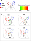
ANA‐positive healthy individuals can be distinguished from SLE patients and ANA‐negative healthy individuals using BLyS, IL‐5, G‐CSF, and IL‐2. A, Random forest models separate SLE patients (red), ANA‐positive healthy individuals (white), and ANA‐negative healthy individuals (blue) by cytokine expression. B, BLyS, IL‐5, G‐CSF, and IL‐2 were used to differentiate ANA‐positive healthy individuals from SLE patients and ANA‐negative healthy controls. Colors indicate the increased cytokine fluorescence intensity, with red as the highest, yellow as average, and green as the lowest. Min = minimum; max = maximum (see Figure 1 for other definitions).
Figure 4
Figure 4
Relative numbers of monocytes and memory B cells are increased in antinuclear antibody−positive (ANA+) healthy individuals. Immunophenotyping flow cytometry was used to determine the total absolute cell number of peripheral blood mononuclear cells (PBMCs)/ml of blood (A) and percentages of PBMCs identified as lymphocytes (B), the T cell subsets CD4+ (C), CD8+ (D), and CD85j+CD4+ (E). The B cell subsets were identified as total (CD3−CD20+CD19+) (F), memory (CD20+CD19+CD27+CD38−) (G), plasmablasts (CD20−CD19+CD27+CD38+) (H), naive (IgD+CD27−CD24−) (I), and transitional (CD27−CD38+CD24+) (J). Frequencies of monocytes (K) and natural killer (NK) cells (CD3−CD56+CD16+) (L) are also shown. Each data point represents an individual subject; horizontal lines show the median and interquartile range. ∗ = P < 0.05; ∗∗ = P < 0.01, by Mann‐Whitney test. ANA− = ANA‐negative; NS = not significant.
Figure 5
Figure 5
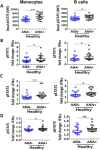
ANA‐positive healthy individuals have increased STAT phosphorylation in B cells and monocytes. Phosphospecific flow cytometry on PBMCs was used to assess basal STAT‐5 phosphorylation (A) and pSTAT‐1 (B), pSTAT‐3 (C), and pSTAT‐5 (D) following interferon‐α (IFNα) stimulation in monocytes and B cells. Each data point represents an individual subject; horizontal lines show the median and interquartile range. ∗ = P < 0.05; ∗∗ = P < 0.01, by Mann‐Whitney test. MFI = mean fluorescence intensity (see Figure 4 for other definitions).

Comment in

Similar articles

Cited by

References

    1. Satoh M, Chan EK, Ho LA, Rose KM, Parks CG, Cohn RD, et al. Prevalence and sociodemographic correlates of antinuclear antibodies in the United States. Arthritis Rheum 2012;64:2319–27. - PMC - PubMed
    1. Parks CG, Miller FW, Satoh M, Chan EK, Andrushchenko Z, Birnbaum LS, et al. Reproductive and hormonal risk factors for antinuclear antibodies (ANA) in a representative sample of U.S. women. Cancer Epidemiol Biomarkers Prev 2014;23:2492–502. - PMC - PubMed
    1. Heinlen LD, McClain MT, Merrill J, Akbarali YW, Edgerton CC, Harley JB, et al. Clinical criteria for systemic lupus erythematosus precede diagnosis, and associated autoantibodies are present before clinical symptoms. Arthritis Rheum 2007;56:2344–51. - PubMed
    1. Arbuckle MR, McClain MT, Rubertone MV, Scofield RH, Dennis GJ, James JA, et al. Development of autoantibodies before the clinical onset of systemic lupus erythematosus. N Engl J Med 2003;349:1526–33. - PubMed
    1. Li QZ, Karp DR, Quan J, Branch VK, Zhou J, Lian Y, et al. Risk factors for ANA positivity in healthy persons. Arthritis Res Ther 2011;13:R38. - PMC - PubMed

MeSH terms

Substances