. 2016 Aug;52(4):484-94.
doi: 10.1007/s11262-016-1325-y. Epub 2016 Apr 8.

Mutations in the C-terminal region affect subcellular localization of crucian carp herpesvirus (CaHV) GPCR

Affiliations

Mutations in the C-terminal region affect subcellular localization of crucian carp herpesvirus (CaHV) GPCR

Jun Wang et al. Virus Genes. 2016 Aug.

Abstract

G protein-coupled receptors (GPCRs) are known as seven transmembrane domain receptors and consequently can mediate diverse biological functions via regulation of their subcellular localization. Crucian carp herpesvirus (CaHV) was recently isolated from infected fish with acute gill hemorrhage. CaHV GPCR of 349 amino acids (aa) was identified based on amino acid identity. A series of variants with truncation/deletion/substitution mutation in the C-terminal (aa 315-349) were constructed and expressed in fathead minnow (FHM) cells. The roles of three key C-terminal regions in subcellular localization of CaHV GPCR were determined. Lysine-315 (K-315) directed the aggregation of the protein preferentially at the nuclear side. Predicted N-myristoylation site (GGGWTR, aa 335-340) was responsible for punctate distribution in periplasm or throughout the cytoplasm. Predicted phosphorylation site (SSR, aa 327-329) and GGGWTR together determined the punctate distribution in cytoplasm. Detection of organelles localization by specific markers showed that the protein retaining K-315 colocalized with the Golgi apparatus. These experiments provided first evidence that different mutations of CaHV GPCR C-terminals have different affects on the subcellular localization of fish herpesvirus-encoded GPCRs. The study provided valuable information and new insights into the precise interactions between herpesvirus and fish cells, and could also provide useful targets for antiviral agents in aquaculture.

Keywords: C-terminal; Crucian carp herpesvirus (CaHV); G protein-coupled receptor (GPCR); N-myristoylation site; Subcellular localization.


Figures

Fig. 1
Fig. 1
Electron micrograph of CaHV. Purified CaHV was negatively stained. The virus particles had icosahedral capsids with protrusions on the surface and had an estimated diameter of 97 nm. Bar = 100 nm
Fig. 2
Fig. 2
Primary sequence, secondary structure, and C-terminal tails of CaHV GPCR. a Nucleotide and amino acid sequence of CaHV GPCR. The putative seven transmembrane domains (I–VII) are shaded and three regions are shown as boxes. b The secondary structure of CaHV GPCR showing the seven membrane spanning domains (I–VII), connecting loops and C-terminal tail with the three key regions. A lysine (K-315, at the position 315), protein kinase C phosphorylation site (PKCs, three residues SSR or Ser-Ser-Arg, at the positions 327–329), and N-myristoylation site (six residues GGGWTR or Gly-Gly-Gly-Trp-Thr-Arg, at the positions 335–340) are shown with boxes, respectively
Fig. 3
Fig. 3
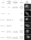
Three key regions of CaHV GPCR play important role in the subcellular localization of GPCR. FHM cells were transfected with expression plasmid pEGFP-GPCR or pEGFP-empty control. At 24 h p.t., cells were fixed for fluorescence observation. Plasmids corresponding to different lengths of CaHV GPCR C-terminal EGFP fusion proteins. Row a. Plasmid names, the plasmids expressing full-length, truncations (T), deletions (D), or substitutions (/) of GPCR C-terminal. The numbers at the plasmid bottom right indicate the position of the C-terminal residues. Row b. Altered aa on number and site (the numbers indicate aa residues). Row c The retaining region (inside the box) present at the C-terminal (the numbers indicate aa residues). Row d The distribution of expressed products in fish FHM cells. Row e The merging fluorescence images of EGFP reporter plasmid expression and subcellular distribution. Bar = 5 μm. The result came from three independent experiments, and at least ten cells were viewed at each time
Fig. 3
Fig. 3
Three key regions of CaHV GPCR play important role in the subcellular localization of GPCR. FHM cells were transfected with expression plasmid pEGFP-GPCR or pEGFP-empty control. At 24 h p.t., cells were fixed for fluorescence observation. Plasmids corresponding to different lengths of CaHV GPCR C-terminal EGFP fusion proteins. Row a. Plasmid names, the plasmids expressing full-length, truncations (T), deletions (D), or substitutions (/) of GPCR C-terminal. The numbers at the plasmid bottom right indicate the position of the C-terminal residues. Row b. Altered aa on number and site (the numbers indicate aa residues). Row c The retaining region (inside the box) present at the C-terminal (the numbers indicate aa residues). Row d The distribution of expressed products in fish FHM cells. Row e The merging fluorescence images of EGFP reporter plasmid expression and subcellular distribution. Bar = 5 μm. The result came from three independent experiments, and at least ten cells were viewed at each time
Fig. 4
Fig. 4
Colocalization of different length GPCR C-terminal with organelle-specific markers. FHM cells were co-transfected organelle-specific marker plasmid with expression plasmid pEGFP-GPCR or pEGFP-empty control. At 24 h p.t., cells were fixed for fluorescence observation. a Colocalization was seen between full-length GPCR and Golgi apparatus (Golgi), and did not colocalize with mitochondria (Mit) or endoplasmic reticulum (ER). b GPCRT-316–349 containing lysine-315 (K-315) colocalized with Golgi, but GPCRT-315–349 without K-315 or empty control did not colocalize with Golgi. The result came from three independent experiments, and at least ten cells were viewed at each time
Fig. 5
Fig. 5
Schematic summary of the effects of C-terminal mutants on the subcellular localization of CaHV GPCR. The variants are represented by four circumstances (dotted line), including K or full length, GGGWTR, SSR and GGGWTR, and control. K or full length: When region K-315 or the intact C-terminal was retained, CaHV GPCR formed aggregates at the nuclear side, and colocalized with the Golgi apparatus and might be transported by Golgi apparatus. GGGWTR: when region GGGWTR was retained but regions K-315 and SSR were missing on the C-terminal, CaHV GPCR showed punctate distribution on the periplasmic side and/or cytoplasm. SSR and GGGWTR: when regions SSR and GGGWTR were both retained, CaHV GPCR exhibited a punctate distribution in the cytoplasm. Control: When empty control or the C-terminal excluding K-315 and GGGWTR were presented, no specific signal was exhibited in the cytoplasm. The distributions of the variants are represented by green (diffusion, uniform, or punctate). CaHV GPCR colocalizing with Golgi apparatus is represented by yellow. The arrows indicate CaHV GPCR and associated proteins might intracellularly transport to peripheral cytoplasm by Golgi apparatus

Similar articles

Cited by

References

    1. Venkatakrishnan AJ, Deupi X, Lebon G, Tate CG, Schertler GF, Babu MM. Molecular signatures of G-protein-coupled receptors. Nature. 2013;494:185–194. doi: 10.1038/nature11896. - DOI - PubMed
    1. Dohlman HG. Thematic mini review series: cell biology of G protein signaling. J. Biological. Chem. 2015;290:6679–6680. doi: 10.1074/jbc.R114.631093. - DOI - PMC - PubMed
    1. Vischer HF, Siderius M, Leurs R, Smit MJ. Herpesvirus-encoded GPCRs: neglected players in inflammatory and proliferative diseases? Nature. Rev. Drug. Discov. 2014;13:123–139. doi: 10.1038/nrd4189. - DOI - PubMed
    1. Rosenbaum DM, Rasmussen SGF, Kobilka BK. The structure and function of G-protein-coupled receptors. Nature. 2009;459:356–363. doi: 10.1038/nature08144. - DOI - PMC - PubMed
    1. Zuo JM, Currin A, Griffin BD, Shannon-Lowe C, Thomas WA, Ressing ME, Wiertz EJHJ, Rowe M. The epstein-barr virus G-protein-coupled teceptor contributes to immune evasion by targeting MHC Class I molecules for degradation. PlosPathog. 2009;5:e1000255. - PMC - PubMed

Substances

LinkOut - more resources