. 2016 Apr 8:15:62.
doi: 10.1186/s12933-016-0377-6.

Nicorandil attenuates carotid intimal hyperplasia after balloon catheter injury in diabetic rats

Affiliations

Nicorandil attenuates carotid intimal hyperplasia after balloon catheter injury in diabetic rats

Ying Qian Zhang et al. Cardiovasc Diabetol. .

Abstract

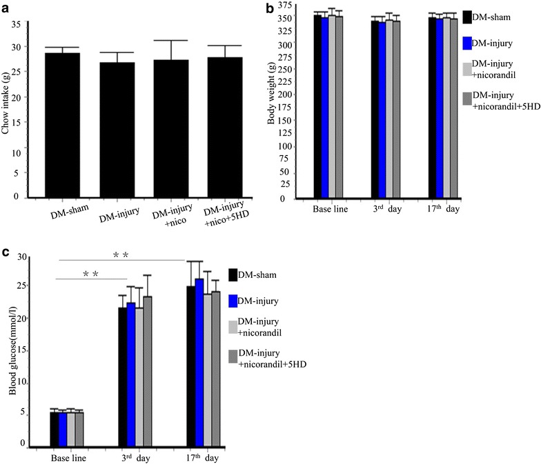
Background: Diabetic patients suffer from undesired intimal hyperplasia after angioplasty. Nicorandil has a trend to reduce the rate of target lesion revascularization. However, whether nicorandil inhibits intimal hyperplasia and the possible mechanisms underlying it remain to be determined. We aimed at assessing the effect of nicorandil on intimal hyperplasia in diabetic rats.

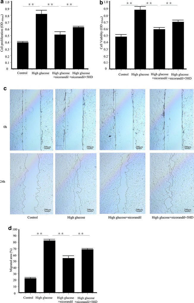
Methods: After intraperitoneal injection of streptozotocin (STZ, 50 mg/kg), balloon injury model was established in carotid arteries of diabetic rats. Rats were randomized to vehicle, nicorandil (15 mg/kg/day) or 5-hydroxydecanoate (5-HD, 10 mg/kg/day), a mitochondrial ATP-sensitive potassium channel (mitoKATP channel)-selective antagonist. Perivascular delivery of εPKC siRNA was conducted to determine the role of εPKC pathway in intimal hyperplasia. In hyperglycemia environment (25 mM glucose), primary culture of vascular smooth muscle cells (VSMCs) were treated with nicorandil or 5-HD. Cell proliferation and cell migration were analyzed.

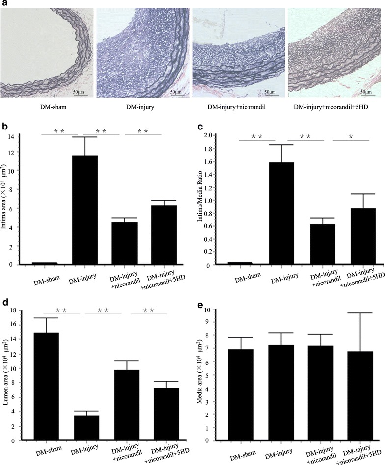
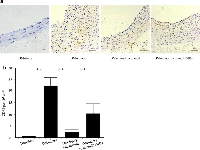
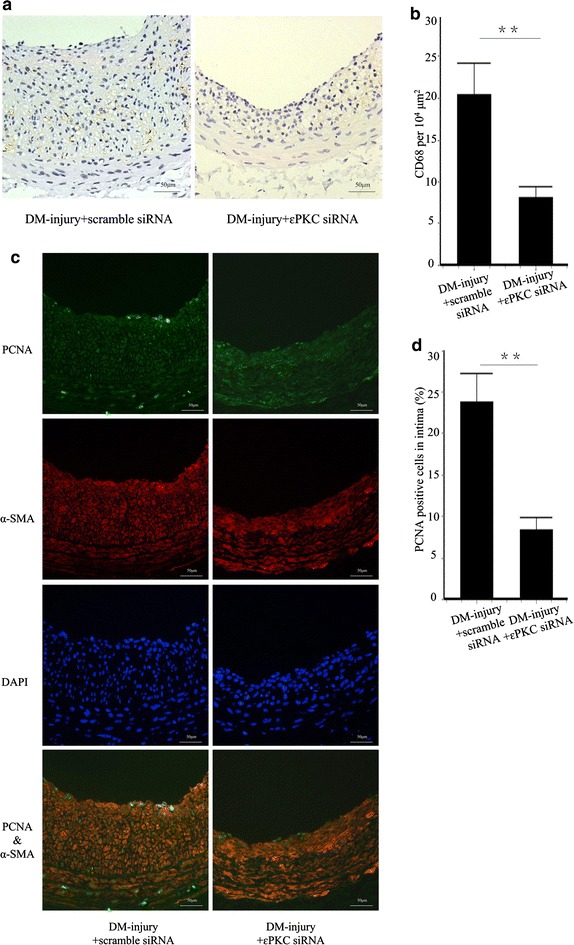
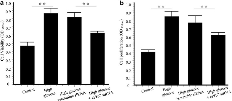
Results: Intimal hyperplasia significantly increased 14 days after balloon injury in diabetic rats (p < 0.01). Nicorandil inhibited intima development, reduced inflammation and prevented cell proliferation in balloon-injured arteries (p < 0.01). The protective effects of nicorandil were reversed by 5-HD (p < 0.05). εPKC was activated in balloon-injured arteries (p < 0.01). Nicorandil inhibited εPKC activation by opening mitoKATP channel. Perivascular delivery of εPKC siRNA inhibited intimal hyperplasia, inflammation and cell proliferation (p < 0.01). High glucose-induced VSMCs proliferation and migration were inhibited by nicorandil. εPKC activation induced by high glucose was also inhibited by nicorandil and that is partially reversed by 5-HD. εPKC knockdown prevented VSMCs proliferation and migration (p < 0.01).

Conclusions: Our study demonstrates that nicorandil inhibits intimal hyperplasia in balloon-injured arteries in diabetic rats. Nicorandil also prevents VSMCs proliferation and migration induced by high glucose. The beneficial effect of nicorandil is conducted via opening mitoKATP channel and inhibiting εPKC activation.

Keywords: ATP-sensitive potassium channel; Diabetes mellitus; Intimal hyperplasia; Nicorandil; Protein kinase C.


Figures

Fig. 1
Fig. 1
Chow intake, body weight and blood glucose in each group. a Chow intakes in different groups. No significant difference was observed among different groups. Bars represent mean ± SE. b Body weight in sham operation group (DM-sham group, n = 8), balloon injury group (DM-injury group, n = 10), nicorandil-treated balloon injury group (DM-injury + nicorandil group, n = 10), and nicorandil and 5-HD-treated group (DM-injury + nicorandil + 5HD group, n = 10). c Blood glucose in DM-sham group, DM-injury group, DM-injury + nicorandil group, and DM-injury + nicorandil + 5HD group. No significant difference was observed among different groups. Blood glucose significantly increased after STZ injection. Bars represent mean ± SE. **p < 0.01
Fig. 2
Fig. 2
Intimal hyperplasia 14 days after balloon injury. a Cross sections of carotid arteries from diabetic rats 14 days after balloon injury. Sections were stained with Elastica van Gieson. b Quantitative analysis of intima area. c Quantitative analysis of intima/media area ratio. d Quantitative analysis of lumen area. e Quantitative analysis of media area in DM-sham group, DM-injury group, DM-injury + nicorandil group, and DM-injury + nicorandil + 5HD group. Nicorandil reduced the intima area and intima/media ratio, and it increased lumen area. The protective effect of nicorandil was significantly blocked by 5-HD. No significant difference was seen in media areas of different groups. Bars represent mean ± SE. **p < 0.01, *p < 0.05
Fig. 3
Fig. 3
Inflammatory cell infiltration in injured carotid arteries. a IHC staining of CD 68 in DM-sham group, DM-injury group, DM-injury + nicorandil group, and DM-injury + nicorandil + 5HD group. b Quantitative analysis of CD 68-posotive cells per 104 μm2. Nicorandil decreased CD 68-positive macrophages in balloon injured arteries. This decrease was reversed by 5-HD. Bars represent mean ± SE. **p < 0.01
Fig. 4
Fig. 4
Cell proliferation in injured carotid arteries. a IF staining of PCNA and α-SMA in DM-sham group, DM-injury group, DM-injury + nicorandil group, and DM-injury + nicorandil + 5HD group. b Quantitative analysis of percentage of PCNA-positive cells in intima. Nicorandil decreased cell proliferation in balloon injured arteries. This decrease was reversed by 5-HD. Bars represent mean ± SE. **p < 0.01
Fig. 5
Fig. 5
Effect of nicorandil on high-glucose-induced VSMCS proliferation and migration. a VSMCs were incubated with high glucose (25 mM) and treated with nicorandil (100 μmol/l) or 5-HD (500 μmol/l). VSMCs proliferation was detected using BrdU proliferation assay kit. 24 h of high glucose induced VSMCs proliferation. Nicorandil inhibited the proliferation and 5-HD partially blocked the effect of nicorandil. b Cell viability was assessed by MTT assay kit. Nicorandil suppressed the increase of cell viability induced by high glucose. 5-HD, a mitoKATP channel-selective antagonist, blocked the effect of nicorandil. c VSMCs migration was assessed by wound healing assay. d Quantitative analysis of percentage of migrated area in control group, high glucose group, high glucose + nicorandil group, and high glucose + nicorandil + 5-HD group. Nicorandil prevented high glucose-induced cell migration by opening mitoKATP channel. Bars represent mean ± SE. **p < 0.01
Fig. 6
Fig. 6
Effect of εPKC on intimal hyperplasia in balloon-injured carotid arteries. a Representative western blots for εPKC translocation in injured carotid artery treated with or without nicorandil or 5-HD. b Quantitative analysis of εPKC translocation. εPKC translocation from the cell soluble (S) to the cell particulate fraction (TS) was observed after balloon injury. This translocation was blocked by nicorandil. The effect of nicorandil was reversed by 5-HD. c Representative western blots for εPKC protein in the injured carotid artery with or without εPKC siRNA. d Quantitative analysis of εPKC protein. εPKC expression in εPKC siRNA delivered group was reduced to 30.41 ± 5.17 % of that in the scramble siRNA delivered group e: Representative cross-section of carotid artery stained with Elastica van Gieson. f Quantitative analysis of of intima/media ratio. g Quantitative analysis of lumen area in DM-injury + scramble siRNA group and DM-injury + εPKC siRNA group. Knockdown of εPKC reduced the intima/media ratio and increased lumen area. Bars represent mean ± SE. **p < 0.01
Fig. 7
Fig. 7
Effect of εPKC on inflammation and cell proliferation in balloon injured carotid arteries. a IHC stain of CD 68 was performed in carotid sections 14 days after balloon injury with or without εPKC siRNA delivery. b Quantitative analysis of CD 68-posotive cells per 104 μm2. c IF staining of PCNA and α-SMA was performed in carotid sections 14 days after balloon injury with or without εPKC siRNA delivery. d Quantitative analysis of percentage of PCNA-positive cells in intima. Labeling of CD 68 and PCNA were significantly lower in εPKC siRNA delivered group than that in scramble siRNA delivered group. Bars represent mean ± SE. ** p < 0.01
Fig. 8
Fig. 8
Effect of εPKC on VSMCs migration induced by high glucose. a Representative western blots for εPKC translocation in VSMCs with or without nicorandil or 5-HD. b Quantitative analysis of εPKC translocation. εPKC translocation was induced by high glucose. Nicorandil inhibited the increase of εPKC translocation by opening mitoKATP channel. c Representative western blots for εPKC protein in VSMCs with or without εPKC siRNA transfection. d Quantitative analysis of εPKC protein. εPKC protein in high-glucose-εPKC siRNA group was reduced to 24.94 ± 7.67 % of that in high-glucose-scramble siRNA group. e VSMCs migration was assessed by wound healing assay. f Quantitative analysis of percentage of migrated area in control group, high glucose + scramble siRNA group, and high glucose + εPKC siRNA group. εPKC siRNA knockdown prevented high glucose-induced cell migration. Bars represent mean ± SE. *p < 0.05, **p < 0.01
Fig. 9
Fig. 9
Effect of εPKC on VSMCs proliferation induced by high glucose. a Cell viability was assessed by MTT assay kit. b VSMCs proliferation was detected using BrdU proliferation assay kit. 24 h of high glucose induced VSMCs proliferation. εPKC knockdown suppressed the increase of cell viability and proliferation induced by high glucose. Bars represent mean ± SE. **p < 0.01

Similar articles

Cited by

References

    1. Srivastava S, Ramana KV, Tammali R, Srivastava SK, Bhatnagar A. Contribution of aldose reductase to diabetic hyperproliferation of vascular smooth muscle cells. Diabetes. 2006;55:901–910. doi: 10.2337/diabetes.55.04.06.db05-0932. - DOI - PMC - PubMed
    1. Fröbert O, Lagerqvist B, Carlsson J, Lindbäck J, Stenestrand U, James SK. Differences in restenosis rate with different drug-eluting stents in patients with and without diabetes mellitus. J Am Coll Cardiol. 2009;53:1660–1667. doi: 10.1016/j.jacc.2009.01.054. - DOI - PubMed
    1. Kawai Y, Hisamatsu K, Matsubara H, Dan K, Akagi S, Miyaji K, et al. Intravenous administration of nicorandil immediately before percutaneous coronary intervention can prevent slow coronary flow phenomenon. Eur Heart J. 2009;30:765–772. doi: 10.1093/eurheartj/ehp077. - DOI - PubMed
    1. Shehata M. Cardioprotective effects of oral nicorandil use in diabetic patients undergoing elective percutaneous coronary intervention. J Interv Cardiol. 2014;27:472–481. doi: 10.1111/joic.12142. - DOI - PubMed
    1. Izumiya Y, Kojima S, Kojima S, Araki S, Usuku H, Matsubara J, et al. Long-term use of oral nicorandil stabilizes coronary plaque in patients with stable angina pectoris. Atherosclerosis. 2011;214:415–421. doi: 10.1016/j.atherosclerosis.2010.11.032. - DOI - PubMed

Publication types

MeSH terms

LinkOut - more resources