. 2016 Sep;68(9):2210-20.
doi: 10.1002/art.39710.

Checkpoints for Autoreactive B Cells in the Peripheral Blood of Lupus Patients Assessed by Flow Cytometry

Affiliations

Checkpoints for Autoreactive B Cells in the Peripheral Blood of Lupus Patients Assessed by Flow Cytometry

Susan Malkiel et al. Arthritis Rheumatol. 2016 Sep.

Abstract

Objective: Antinuclear antibodies (ANAs) are diagnostic in several autoimmune disorders, yet the failure to achieve B cell tolerance in these diseases is still poorly understood. Although secreted ANAs detected by an indirect immunofluorescence assay are the gold standard for autoreactivity, there has been no convenient assay with which to measure the frequency of circulating B cells that recognize nuclear antigens (ANA+ B cells) in patients. The aim of this study was to generate an assay to easily identify these B cells and to examine its utility in a study of autoreactive B cells in systemic lupus erythematosus (SLE).

Methods: We developed and validated a novel flow cytometry-based assay that identifies ANA+ B cells using biotinylated nuclear extracts, and utilized it to examine B cell tolerance checkpoints in peripheral blood mononuclear cells obtained from SLE patients and healthy controls.

Results: We observed progressive selection against ANA+ B cells as they matured from transitional to naive to CD27+IgD- and CD27+IgD+ memory cells in both healthy subjects and SLE patients; however, ANA+ naive B cells in SLE patients were not anergized to the same extent as in healthy individuals. We also showed that anergy induction is restored in SLE patients treated with belimumab, an inhibitor of BAFF.

Conclusion: This assay will enable studies of large populations to identify potential genetic or environmental factors affecting B cell tolerance checkpoints in healthy subjects and patients with autoimmune disease and permit monitoring of the B cell response to therapeutic interventions.


Figures

Figure 1
Figure 1
Identification of B cells that recognize nuclear antigens (ANA+ B cells) by flow cytometry, and validation of the assay. A, Composition of HeLa nuclear extracts analyzed by agarose gel electrophoresis (left), sodium dodecyl sulfate–polyacrylamide gel electrophoresis (middle, left), and immunoblotting (right). B, Gating strategy for flow cytometry–based antinuclear antibody (ANA) assay, along with strategy used to sort single ANA+ and ANA− naive and transitional B cells for cloning of immunoglobulin genes. After gating on lymphocytes and excluding doublets by FSC and SSC (plots not shown), lymphocytes were further resolved into non-B cell (CD19−CD3+CD14+CD16+) and B cell (CD19+CD3−CD14−CD16−) populations. ANA+ B cells were identified using non-B cells as a negative control. C, Representative immunofluorescence staining patterns, on HEp-2–coated slides, of purified recombinant antibodies derived from sorted ANA+ (a–e) and ANA− (f) B cells from healthy controls, showing a variety of staining patterns: homogeneous (a), speckled (b), rim (c), centromere (d), and nuclear/cytoplasmic (e). D, ANA binding of recombinant antibodies from sorted ANA+ (n = 23) and ANA− (n = 17) B cells as measured by ANA enzyme-linked immunosorbent assay. Each data point represents the mean absorbance value of triplicates for each antibody sample. The red triangle in the ANA− group represents the negative control antibody B1. The dotted line represents the cutoff OD for ANA reactivity. Experiments were performed at least twice.
Figure 2
Figure 2
Antinuclear reactivity in peripheral B cell subsets from systemic lupus erythematosus (SLE) patients and healthy control (HC) subjects. A, The gating strategy used to identify different B cell subsets. After resolving lymphocytes into non-B cells and B cells (as described in Figure 1B), CD27 and IgD staining was used to further define B cells into CD27+IgD+ memory, CD27+IgD− memory, double-negative (DN), and non-memory (CD27− IgD+) subsets. CD10 and CD38 expression was used to distinguish transitional (CD38highCD10high) and naive (CD38intermediateCD10low/−) B cells in the non-memory B cell subset. B, Zebra plots for a representative SLE patient, a healthy control subject, and a belimumab-treated SLE patient. B cells recognizing nuclear antigens (ANA+ B cells) were identified within each B cell subset. Non-B cells were used as a negative control to set the antinuclear antibody (ANA) gate. C, Scatterplots depicting the frequency of ANA+ B cells within each B cell subset in SLE patients (n = 37), healthy controls (n = 33), and belimumab-treated SLE patients (n = 9). Each data point represents the frequency in an individual subject; horizontal lines show the mean. P values were determined by 2-tailed Mann-Whitney test. NS = not significant.
Figure 3
Figure 3
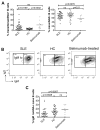
Frequency of anergic ANA+ naive B cells in SLE patients and healthy control subjects. A, Scatterplots depicting the proportions of ANA+ B cells that are transitional and naive in SLE patients (n = 37), healthy controls (n = 33), and belimumab-treated SLE patients (n = 9). Total ANA+ B cells were first identified as described in Figure 1B and subsequently gated on the B cell subsets described in Figure 2. B, Representative contour plots depicting IgMlow cells within the ANA+ naive B cell compartment of an SLE patient, a healthy control, and a belimumab-treated SLE patient. The gate for IgMlow B cells was determined individually for each sample, using non-B cells as a guide. C, Percentages of IgMlow B cells in the ANA+ naive B cell compartments of patients with SLE, healthy controls, and belimumab-treated SLE patients. P values were determined by 2-tailed Mann-Whitney test. In A and C, each data point represents the frequency in an individual subject; horizontal lines show the mean. Lo = low (see Figure 2 for other definitions).

Similar articles

Cited by

References

    1. Wardemann H, Yurasov S, Schaefer A, Young JW, Meffre E, Nussenzweig MC. Predominant autoantibody production by early human B cell precursors. Science. 2003;301:1374–7. - PubMed
    1. Mahler M, Meroni PL, Bossuyt X, Fritzler MJ. Current concepts and future directions for the assessment of autoantibodies to cellular antigens referred to as anti-nuclear antibodies. J Immunol Res. 2014;2014:315179. - PMC - PubMed
    1. Yurasov S, Hammersen J, Tiller T, Tsuiji M, Wardemann H. B-cell tolerance checkpoints in healthy humans and patients with systemic lupus erythematosus. Ann N Y Acad Sci. 2005;1062:165–74. - PubMed
    1. Yurasov S, Wardemann H, Hammersen J, Tsuiji M, Meffre E, Pascual V, et al. Defective B cell tolerance checkpoints in systemic lupus erythematosus. J Exp Med. 2005;201:703–11. - PMC - PubMed
    1. Tiller T, Meffre E, Yurasov S, Tsuiji M, Nussenzweig MC, Wardemann H. Efficient generation of monoclonal antibodies from single human B cells by single cell RT-PCR and expression vector cloning [published erratum appears in J Immunol Methods 2008;334:142] J Immunol Methods. 2008;329:112–24. - PMC - PubMed

Publication types