Review
. 2016 Apr;137(4):984-997.
doi: 10.1016/j.jaci.2016.02.004.

Molecular and cellular mechanisms of food allergy and food tolerance

Affiliations
Review

Molecular and cellular mechanisms of food allergy and food tolerance

R Sharon Chinthrajah et al. J Allergy Clin Immunol. 2016 Apr.

Abstract

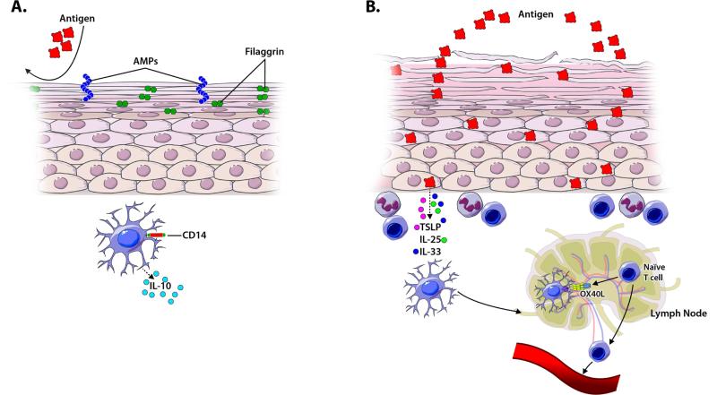
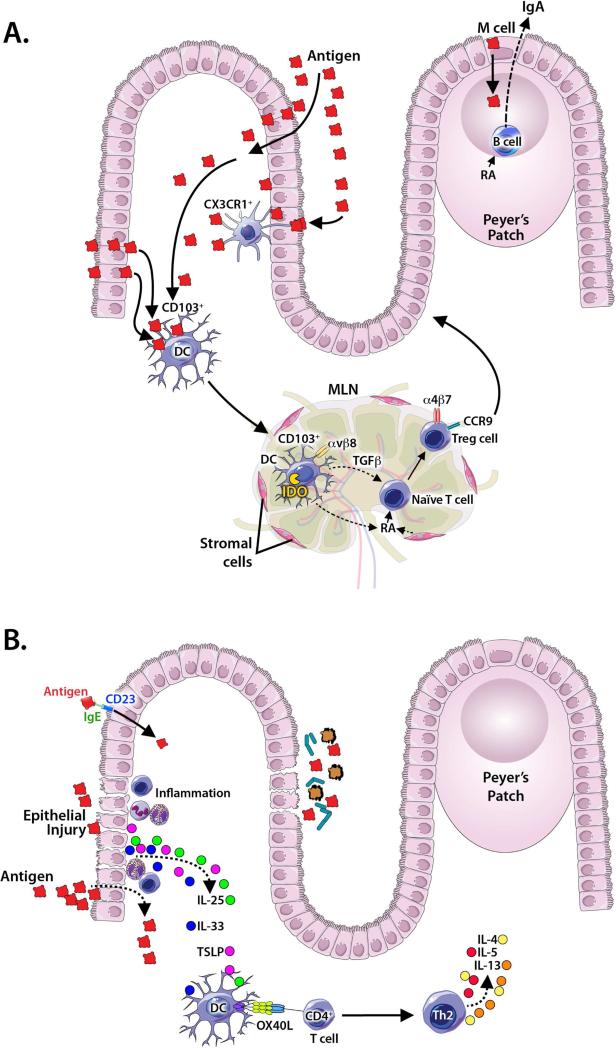
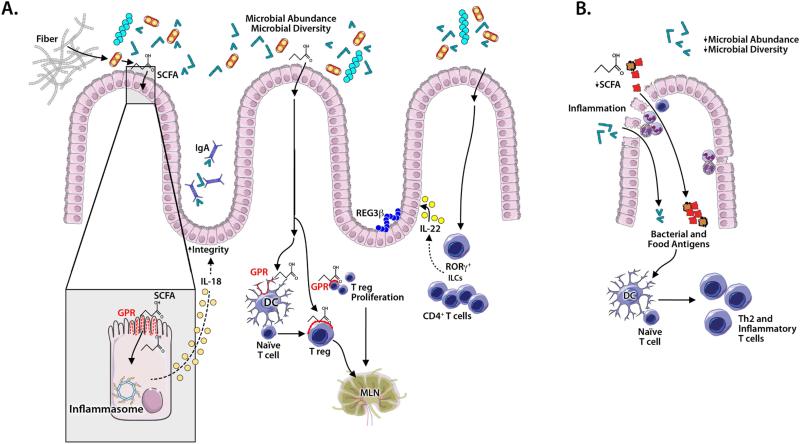
Ingestion of innocuous antigens, including food proteins, normally results in local and systemic immune nonresponsiveness in a process termed oral tolerance. Oral tolerance to food proteins is likely to be intimately linked to mechanisms that are responsible for gastrointestinal tolerance to large numbers of commensal microbes. Here we review our current understanding of the immune mechanisms responsible for oral tolerance and how perturbations in these mechanisms might promote the loss of oral tolerance and development of food allergies. Roles for the commensal microbiome in promoting oral tolerance and the association of intestinal dysbiosis with food allergy are discussed. Growing evidence supports cutaneous sensitization to food antigens as one possible mechanism leading to the failure to develop or loss of oral tolerance. A goal of immunotherapy for food allergies is to induce sustained desensitization or even true long-term oral tolerance to food allergens through mechanisms that might in part overlap with those associated with the development of natural oral tolerance.

Keywords: Food allergy; basophils; dendritic cells; desensitization; immunotherapy; mast cells; microbiome; regulatory T cells; sensitization; tolerance.


Figures

Figure 1
Figure 1. A model of how the gut promotes tolerance or sensitization
Protein antigens (Ags) pass through the epithelial barrier via multiple mechanisms including capture by transluminal processes of CX3CR1+ cells. CD103+ dendritic cells (DCs) then capture Ag, migrate to the mesenteric lymph nodes (MLNs), and present Ag to naïve T cells. In tolerance (A), this interaction promotes the generation of Tregs via 1) production of retinoic acid (RA) by MLN, 2) DC expression of IDO, 3) DC secretion of TGFβ, and 4) DC upregulation of αvβ8 to activate latent TGFβ. Gut homing receptors, CCR9 and α4β7, are upregulated on newly formed Tregs. RA and DC interactions also stimulate differentiation of IgA producing B cells. In sensitization (B), epithelial disruption allows increased antigen penetration, and promotes production/release of epithelial cytokines (IL-33, TSLP and IL-25) that upregulate OX40 ligand (OX40L) on DCs. DCs then promote differentiation of naïve T cells to Th2 cells producing cytokines that recruit eosinophils (IL-5) and promote IgE class switching in B cells (IL-4 and IL-13). IgE may facilitate Ag uptake through CD23.
Figure 2
Figure 2. A model of how the skin promotes tolerance or sensitization
Keratinocyte differentiation, an intact uppermost stratum corneum layer with epidermal proteins that maintain barrier function (eg. filaggrin), antimicrobial peptides (AMPs), and tolerogenic APCs, such as CD14+ DCs producing IL-10, are important for promoting tolerance in the skin barrier (A). Loss of barrier function in the stratum corneum, allowing increased Ag penetration, can occur as a result of genetically-determined defects in factors necessary for keratinocyte differentiation (eg. mutations in filaggrin) or as a result of inflammatory skin diseases (eg. atopic dermatitis). In response to injury, activation by microbial or food antigens, or inflammatory signals TSLP, IL-33 and/or IL-25, produced by keratinocytes, can upregulate OX40L on APCs to promote Th2 differentiation (B).
Figure 3
Figure 3. Microbial mechanisms contributing to oral tolerance and allergic sensitization in the colon
Microbial diversity and abundance promote tolerance (A). Microbes ferment fiber to produce short chain fatty acids (SCFAs) that bind G-protein coupled receptors (GPRs) on: 1) intestinal epithelial cells (IECs) to activate inflammasome production of IL-18 that promotes epithelial barrier integrity; 2) dendritic cells (DCs) to drive naïve T cells to become Tregs; 3) Tregs to induce proliferation. Additionally, SCFAs promote acetylation of histone H3 to preserve or induce FoxP3+ Tregs. Microbe induced IL-22 production by RORγt+ innate lymphocytes and CD4+ T cells promotes barrier integrity and IEC synthesis of antimicrobial peptides and mucus. “Tolerogenic” colonic DCs and lymphocytes likely migrate to mesenteric lymph nodes. In allergic sensitization (B), changes in microbial abundance and diversity (eg, after antibiotic exposure) decrease SCFA, IL-18 and IL-22 levels, compromising epithelial integrity, thereby facilitating epithelial passage of microbial and food antigens. DC activation promotes inflammation, the development of Th2 cell-associated immune responses (including production of allergen-specific IgE antibodies), and allergic sensitization.

References

    1. Moog F. The lining of the small intestine. Sci Am. 1981;245:154–8. 60, 62. et passiom. - PubMed
    1. Brandtzaeg P. Development and basic mechanisms of human gut immunity. Nutr Rev. 1998;56:S5–18. - PubMed
    1. Lozupone CA, Stombaugh JI, Gordon JI, Jansson JK, Knight R. Diversity, stability and resilience of the human gut microbiota. Nature. 2012;489:220–30. - PMC - PubMed
    1. Mestecky J, McGhee JR. Immunoglobulin A (IgA): molecular and cellular interactions involved in IgA biosynthesis and immune response. Adv Immunol. 1987;40:153–245. - PubMed
    1. van der Heijden PJ, Stok W, Bianchi AT. Contribution of immunoglobulin-secreting cells in the murine small intestine to the total 'background' immunoglobulin production. Immunology. 1987;62:551–5. - PMC - PubMed

Publication types

MeSH terms