

Cerebrospinal Fluid Biomarkers of Simian Immunodeficiency Virus Encephalitis : CSF Biomarkers of SIV Encephalitis
- PMID: 27059917
- PMCID: PMC4871628
- DOI: 10.1007/s11481-016-9666-9
Cerebrospinal Fluid Biomarkers of Simian Immunodeficiency Virus Encephalitis : CSF Biomarkers of SIV Encephalitis
Abstract
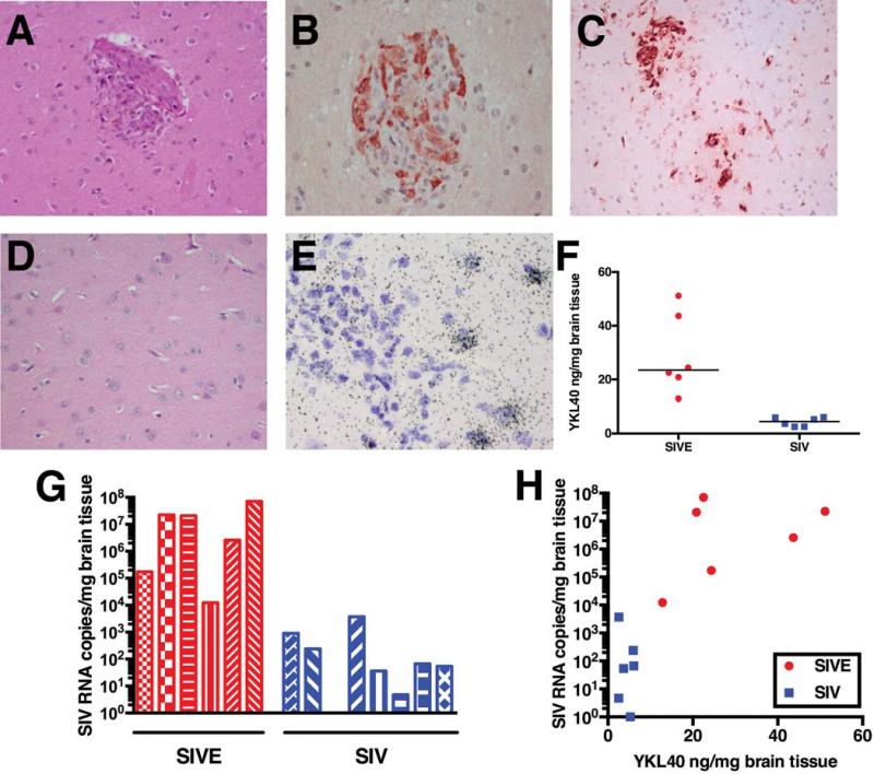
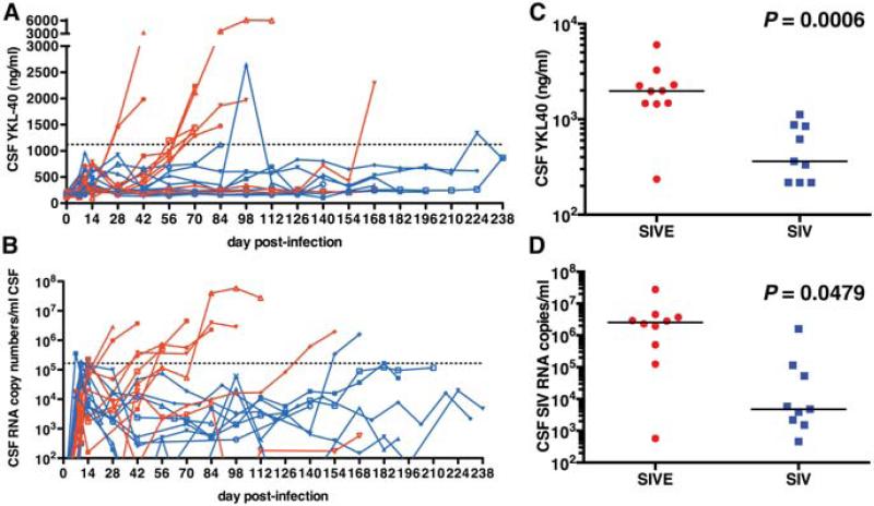
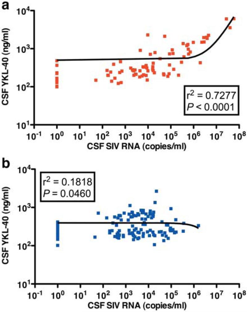
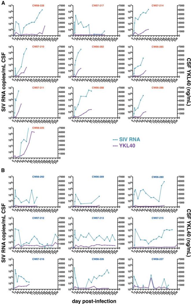
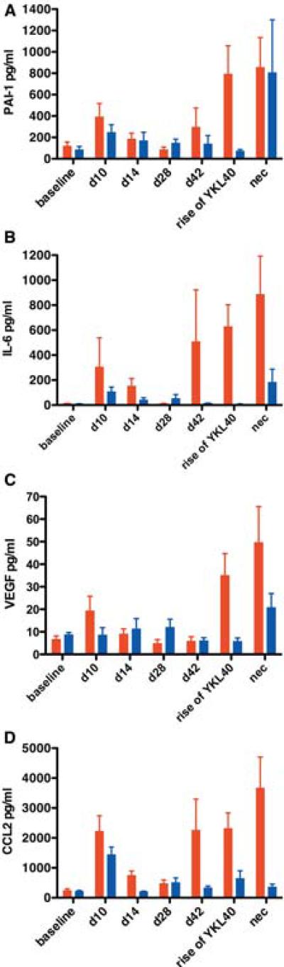
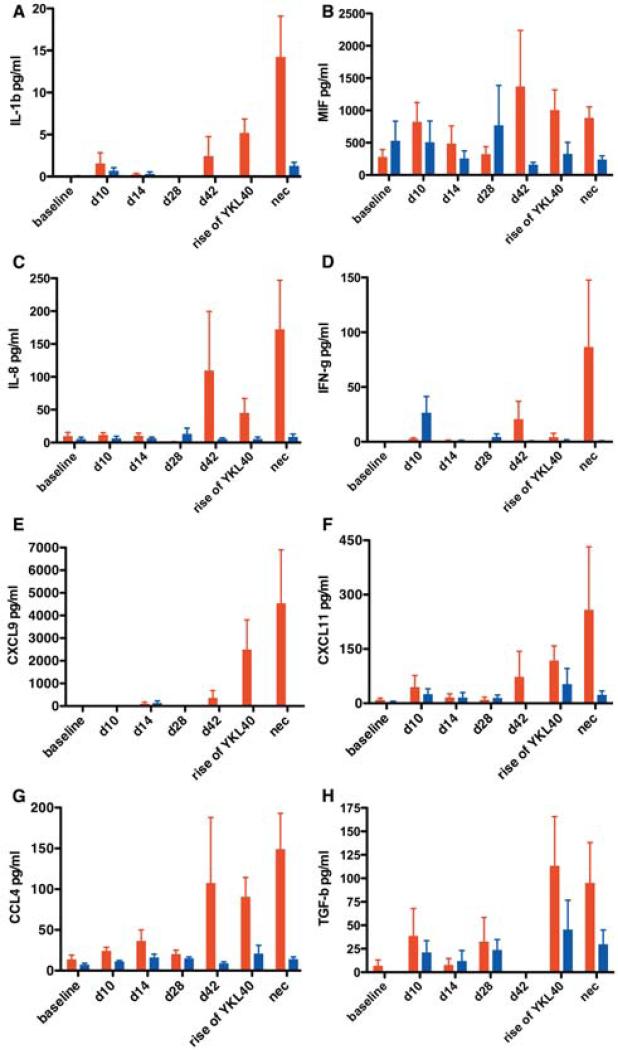
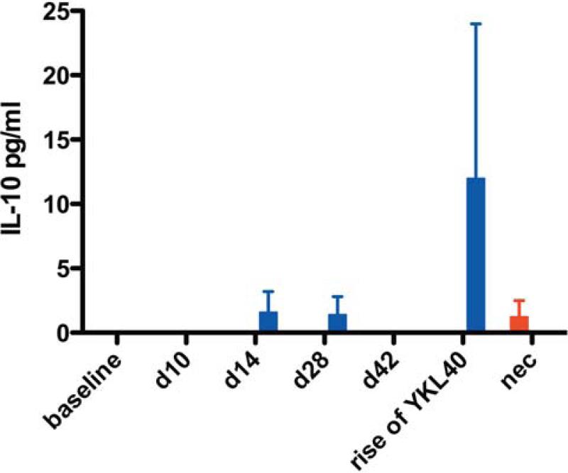
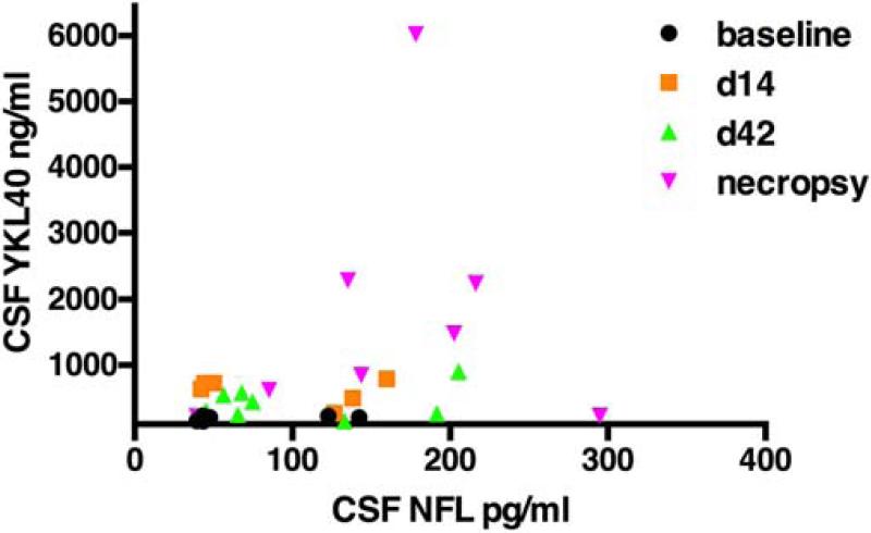
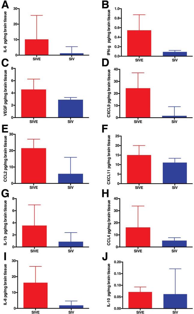
Antiretroviral therapy has led to increased survival of HIV-infected patients but also increased prevalence of HIV-associated neurocognitive disorders. We previously identified YKL40 as a potential cerebrospinal fluid (CSF) biomarker of lentiviral central nervous system (CNS) disease in HIV-infected patients and in the macaque model of HIV encephalitis. The aim of this study was to define the specificity and sensitivity along with the predictive value of YKL40 as a biomarker of encephalitis and to assess its relationship to CSF viral load. CSF YKL40 and SIV RNA concentrations were analyzed over the course of infection in 19 SIV-infected pigtailed macaques and statistical analyses were performed to evaluate the relationship to encephalitis. Using these relationships, CSF alterations of 31 neuroimmune markers were studied pre-infection, during acute and asymptomatic infection, at the onset of encephalitis, and at necropsy. YKL40 CSF concentrations above 1122 ng/ml were found to be a specific and sensitive biomarker for the presence of encephalitis and were highly correlated with CSF viral load. Macaques that developed encephalitis had evidence of chronic CNS immune activation during early, asymptomatic, and end stages of infection. At the onset of encephalitis, CSF demonstrated a rise of neuroimmune markers associated with macrophage recruitment, activation and interferon response. CSF YKL40 concentration and viral load are valuable biomarkers to define the onset of encephalitis. Chronic CNS immune activation precedes the development of encephalitis while some responses suggest protection from CNS lentiviral disease.
Keywords: Biomarker; Cerebrospinal fluid; Encephalitis; Human immunodeficiency virus; Simian immunodeficiency virus; YKL40.
Figures
Similar articles
-
High viral load in the cerebrospinal fluid and brain correlates with severity of simian immunodeficiency virus encephalitis.J Virol. 1999 Dec;73(12):10480-8. doi: 10.1128/JVI.73.12.10480-10488.1999. J Virol. 1999. PMID: 10559366 Free PMC article.
-
YKL-40, a marker of simian immunodeficiency virus encephalitis, modulates the biological activity of basic fibroblast growth factor.Am J Pathol. 2008 Jul;173(1):130-43. doi: 10.2353/ajpath.2008.080045. Epub 2008 Jun 13. Am J Pathol. 2008. PMID: 18556781 Free PMC article.
-
Neuroprotective and anti-human immunodeficiency virus activity of minocycline.JAMA. 2005 Apr 27;293(16):2003-11. doi: 10.1001/jama.293.16.2003. JAMA. 2005. PMID: 15855434
-
Paving the path to HIV neurotherapy: Predicting SIV CNS disease.Eur J Pharmacol. 2015 Jul 15;759:303-12. doi: 10.1016/j.ejphar.2015.03.018. Epub 2015 Mar 24. Eur J Pharmacol. 2015. PMID: 25818747 Free PMC article. Review.
-
An SIV/macaque model targeted to study HIV-associated neurocognitive disorders.J Neurovirol. 2018 Apr;24(2):204-212. doi: 10.1007/s13365-017-0582-4. Epub 2017 Oct 3. J Neurovirol. 2018. PMID: 28975505 Free PMC article. Review.
Cited by
-
CSF Inflammation Markers Associated with Asymptomatic Viral Escape in Cerebrospinal Fluid of HIV-Positive Individuals on Antiretroviral Therapy.Viruses. 2023 Aug 29;15(9):1829. doi: 10.3390/v15091829. Viruses. 2023. PMID: 37766236 Free PMC article.
-
Advances in SIV/SHIV Non-Human Primate Models of NeuroAIDS.Pathogens. 2021 Aug 12;10(8):1018. doi: 10.3390/pathogens10081018. Pathogens. 2021. PMID: 34451482 Free PMC article. Review.
-
The Role of Adipokines in the Pathologies of the Central Nervous System.Int J Mol Sci. 2023 Sep 28;24(19):14684. doi: 10.3390/ijms241914684. Int J Mol Sci. 2023. PMID: 37834128 Free PMC article. Review.
References
-
- Airoldi M, Bandera A, Trabattoni D, Tagliabue B, Arosio B, Soria A, Rainone V, Lapadula G, Annoni G, Clerici M, Gori A. Neurocognitive impairment in HIV-infected naive patients with advanced disease: the role of virus and intrathecal immune activation. Clin Dev Immunol. 2012;2012:467154. - PMC - PubMed
-
- An SF, Groves M, Gray F, Scaravilli F. Early entry and widespread cellular involvement of HIV-1 DNA in brains of HIV-1 positive asymptomatic individuals. J Neuropathol Exp Neurol. 1999;58:1156–1162. - PubMed
-
- Ancuta P, Bakri Y, Chomont N, Hocini H, Gabuzda D, Haeffner-Cavaillon N. Opposite effects of IL-10 on the ability of dendritic cells and macrophages to replicate primary CXCR4-dependent HIV-1 strains. J Immunol. 2001;166:4244–4253. - PubMed
-
- Andreasson U, Portelius E, Andersson ME, Blennow K, Zetterberg H. Aspects of beta-amyloid as a biomarker for Alzheimer's disease. Biomark Med. 2007;1:59–78. - PubMed
Publication types
MeSH terms
Substances
Grants and funding
LinkOut - more resources
Full Text Sources
Other Literature Sources
