. 2016 Jun 10;291(24):12732-12746.
doi: 10.1074/jbc.M115.704734. Epub 2016 Apr 8.

Extracellular Regulation of Bone Morphogenetic Protein Activity by the Microfibril Component Fibrillin-1

Affiliations

Extracellular Regulation of Bone Morphogenetic Protein Activity by the Microfibril Component Fibrillin-1

Alexander P Wohl et al. J Biol Chem. .

Abstract

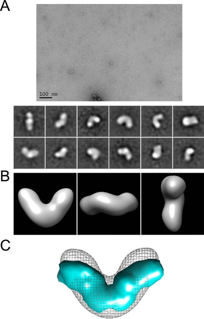
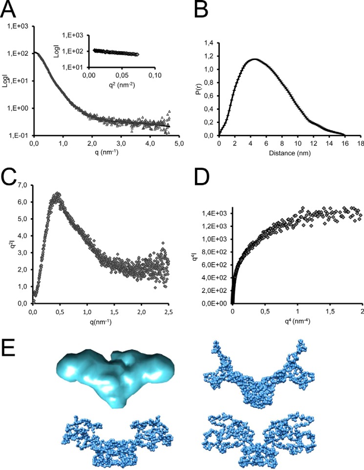
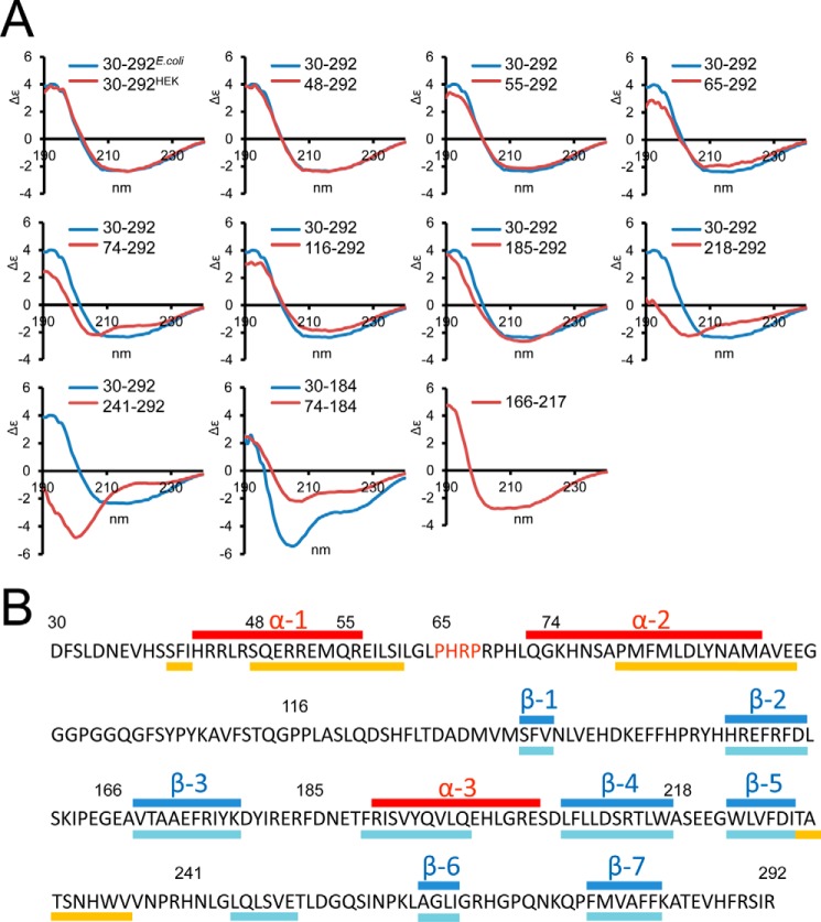
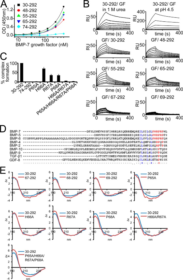
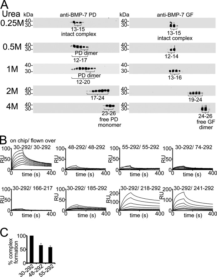
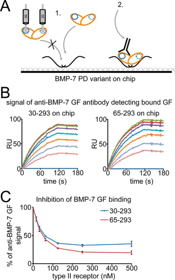
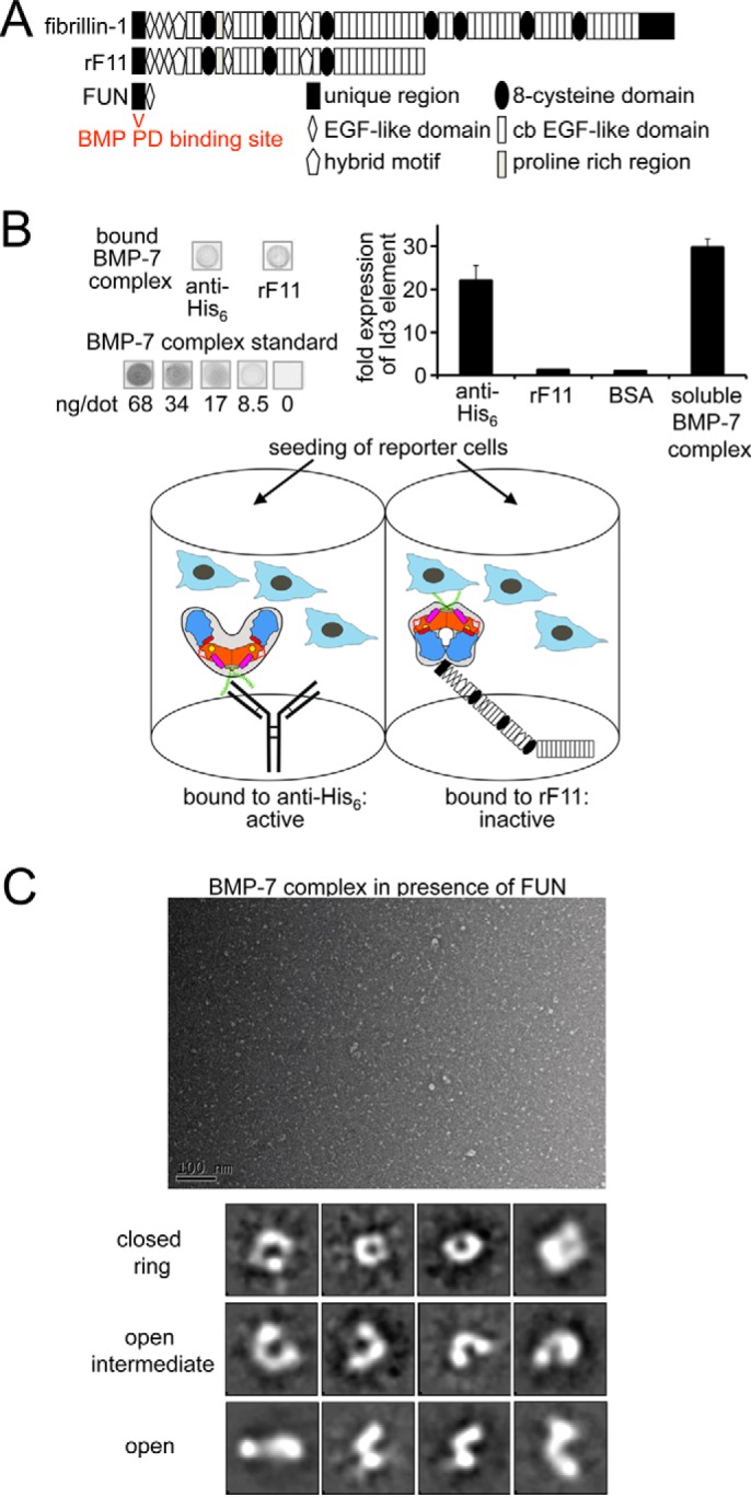
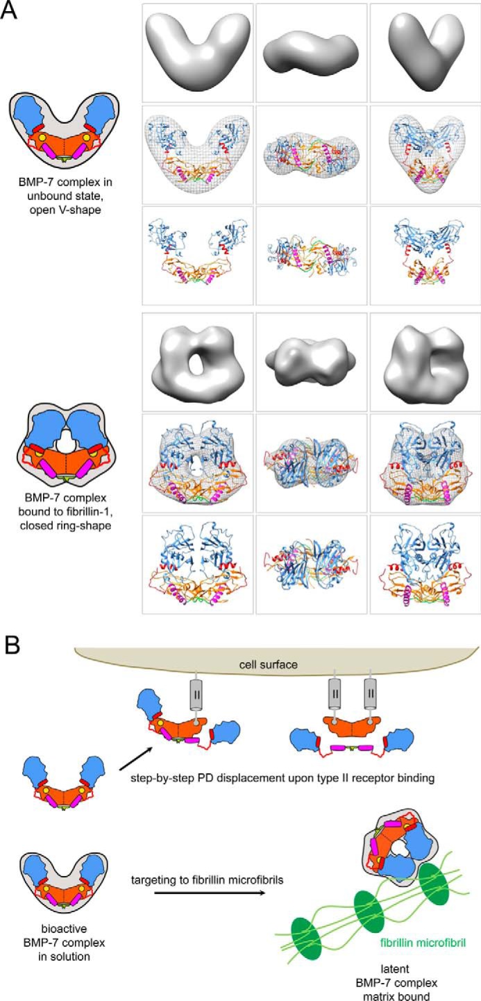
Since the discovery of bone morphogenetic proteins (BMPs) as pluripotent cytokines extractable from bone matrix, it has been speculated how targeting of BMPs to the extracellular matrix (ECM) modulates their bioavailability. Understanding these processes is crucial for elucidating pathomechanisms of connective tissue disorders characterized by ECM deficiency and growth factor dysregulation. Here, we provide evidence for a new BMP targeting and sequestration mechanism that is controlled by the ECM molecule fibrillin-1. We present the nanoscale structure of the BMP-7 prodomain-growth factor complex using electron microscopy, small angle x-ray scattering, and circular dichroism spectroscopy, showing that it assumes an open V-like structure when it is bioactive. However, upon binding to fibrillin-1, the BMP-7 complex is rendered into a closed ring shape, which also confers latency to the growth factor, as demonstrated by bioactivity measurements. BMP-7 prodomain variants were used to map the critical epitopes for prodomain-growth factor and prodomain-prodomain binding. Together, these data show that upon prodomain binding to fibrillin-1, the BMP-7 complex undergoes a conformational change, which denies access of BMP receptors to the growth factor.

Keywords: bone morphogenetic protein (BMP); electron microscopy (EM); extracellular matrix; fribrillin; growth factor; signal transduction; small-angle X-ray scattering (SAXS).


Figures

FIGURE 1.
FIGURE 1.
Affinity purification of recombinant proteins used in this study. A, Coomassie Brilliant Blue-stained SDS-polyacrylamide quality control gels of recombinantly expressed and affinity-purified BMP-7 PD variants and proteins representing the fibrillin-1 N terminus. B (left), size exclusion chromatogram of the BMP-7 PD-GF complex after chelating chromatography utilizing the His6 tag placed at the N terminus of the PD. The chromatogram shows the BMP-7 PD-GF complex mainly eluting in one peak. Right, Coomassie Brilliant Blue-stained SDS-polyacrylamide gel showing the purity of the peak fraction. C, Coomassie Brilliant Blue-stained SDS-polyacrylamide gels showing successful separation of the GF from the PD. The separation was performed as described previously (20). BMP-7 complex was separated into BMP-7 PD (34 kDa) and GF dimer (31 kDa) after dialysis into 8 m urea followed by chelating chromatography, where the PD was bound to the affinity column, and the GF was obtained in the flow-through.
FIGURE 2.
FIGURE 2.
Three-dimensional EM and solution SAXS models of BMP-7 PD-GF complex. Three-dimensional structure of BMP-7 complex was generated using TEM. A (top), representative electron micrograph of BMP-7 complex molecules (scale bar, 100 nm); bottom, 12 images selected from 140 class sum images of 9,000 particles that represent different views of BMP-7 complex (box size = 29.4 × 29.4 nm). B, class sum images were used to generate a three-dimensional TEM model of BMP-7 complex with 2-fold symmetry using angular reconstitution. C, superimposition of the BMP-9 complex structure (33) at 20 Å with the determined BMP-7 EM envelope suggests that the angle between the boomerang arms may be wider in BMP-9.
FIGURE 3.
FIGURE 3.
SAXS data collected for BMP-7 complex. A, x-ray scattering profile of BMP-7 complex showing intensity as a function of q (gray triangles) and Gnom fit to the data (black line). Inset, Guinier plot showing Rg of 4.8 nm. B, P(r) plot showing Dmax of 16 nm. C, Kratky plot showing profile typical of a folded protein. D, plateau in the Porod-Debye plot indicative of a non-flexible protein. E, ab initio models generated from SAXS data using GASBOR with 2-fold symmetry (blue); the averaged model is shown along with three representative models.
FIGURE 4.
FIGURE 4.
Secondary structure analysis of BMP-7 PD. A, CD measurements of N-terminal BMP-7 PD truncation variants (red) in comparison with full-length BMP-7 PD (30–292, blue). Bottom panel, middle, truncation of the first 43 residues in a shorter variant covering the first 184 N-terminal residues (red) results in significant reduction of the α-helical peak at 209 nm compared with the control (blue). Construct 166–217 suggests the existence of a third α-helix within this region. Secondary structure percentage calculated from these CD curves is listed in Tables 1 and 2. B, secondary structure prediction based on CD measurements shows the position of α-helices (red). The position of β-sheets (blue) was guided by predicted secondary structure elements (PSIPRED) marked below the sequence (yellow, α-helical regions; light blue, β-sheets).
FIGURE 5.
FIGURE 5.
Identification of the GF binding motif within the BMP-7 PD. A, solid phase binding ELISA-style assays with immobilized BMP-7 PD truncation variants and GF in solution. B, SPR binding studies of BMP-7 N-terminal PD truncation variants and BMP-7 GF. Top panel, GF binding to the PD was robust to the presence of 1 m urea and pH reduction to 4.5 (full-length PD(30–292) immobilized, GF in solution). Bottom panels, GF was immobilized, and PD variants were injected in solution. C, BMP-7 reconstitution after dialysis of PD variants and GF. Successful reconstitution was monitored in a sandwich ELISA using an anti-His6 antibody against the C-terminal His6 tag on the PD as capture and a polyclonal anti-BMP-7 GF antibody as detector. Error bars, S.D. from three independent experiments. D, sequence alignment using ClustalW identifies the 65PHRP68 motif (red) as conserved within the BMP-5, -6, -7 subgroup of the TGF-β superfamily. Blue, predicted Ile58-Leu59-Leu62-Leu64 GF binding motif (35). E, CD spectra of systematic truncation variants between Arg67 and Pro69. Deletion of Pro65-His66 results in a significant increase of α-helical content of 8% when compared with the 65–292 PD variant (Table 1). This increase returned to normal levels upon stepwise N-terminal truncation of the subsequent two residues. The point mutation P65A resulted in a 4% increase of α-helical content (Table 1), whereas H66A or P68A resulted in no or little change. Additional mutation of the subsequent three residues resulted in no additional change in α-helical content in the quadruple mutant variant P65A/H66A/R67A/P68A (Table 1).
FIGURE 6.
FIGURE 6.
BMP-7 PD interacts with itself. A, dialysis of BMP-7 complex into 0.25–4 m urea reveals the presence of PD dimers monitored by velocity sedimentation experiments using 5–20% sucrose gradients. Each gradient was collected in 28 fractions (fraction 1 at 5% and fraction 28 at 20% sucrose) and subjected to Western blotting analysis for BMP-7 complex components. Western blots were incubated with anti-BMP-7 PD antibody first, stripped, and subsequently re-incubated with anti-BMP-7 GF antibody. B, SPR sensorgrams of self-interaction studies with BMP-7 full-length PD, 48–292, and 55–292, respectively, of immobilized full-length PD and PD variants representing the C-terminal end. C, BMP-7 complex reconstitution is affected by 30% upon deletion of the N-terminal PD self-interaction site. Error bars, mean ± S.D. from three independent experiments.
FIGURE 7.
FIGURE 7.
The 65PHRP68 motif within the N-terminal region of the BMP-7 PD is required for competition with the BMP type II receptor for GF binding. A, scheme of the experimental set-up. Full-length BMP-7 PD (residues 30–292) and the N-terminal truncation variant 65–292 were immobilized, and 100 nm BMP-7 GF was injected in the presence of 0–500 nm BMPRII receptor extracellular domain onto the chip first, followed by a second injection of 100 nm mAb anti-BMP-7 GF antibody to detect bound GF (all injections were in HBS-EP buffer). B, sensorgrams of 100 nm injected mAb anti-BMP-7 GF antibody to detect bound GF. C, increasing amounts of receptor resulted in comparable inhibition of GF binding to both immobilized PD variants, suggesting that the presence of the 65PHRP68 motif in 65–292 is responsible for PD competition with the type II receptor for the same binding site on the GF. Error bars, S.D. from three independent experiments. The schematic shows GF (orange) and type II receptor (blue) binding sites.
FIGURE 8.
FIGURE 8.
Binding to fibrillin-1 induces a conformational change of the BMP-7 complex, resulting in GF inhibition. A, domain structure of fibrillin-1 and used variants. B, BMP activity assay with BMP-7 complex captured via PD interactions, mAb against the N-terminal His6 tag, or the N-terminal half of fibrillin-1 (rF11). C2C12 cells were seeded onto immobilized BMP-7 complex, and Id3 expression was measured as a read-out for BMP activity. Shown is dot blotting analysis of stripped BMP-7 complex by comparison with a diluted series of dots containing BMP-7 complex at known concentrations. The schematic depicts the different ways BMP-7 PD-GF complex is presented to the reporter cells. Antibody capture of the N-terminally placed His6 tag on the PD (green) does not affect bioactivity; however, binding of fibrillin-1 within the PD (blue) induces a conformational change into a ring shape that confers latency. Orange, GF dimer; yellow circle, type II receptor binding site; green, N-terminal His6 tag; magenta, α1-helix; red, α2-helix; red, stretch connecting α1- and α2-helix containing the 65PHRP68 motif; light blue, C-terminal portion of BMP-7 PD. C, dialyzing the small fibrillin-1 N-terminal fragment FUN to BMP-7 complex resulted in the formation of ring shapes and open intermediates that were absent in the BMP-7 complex-only sample (Fig. 2A). Shown are a representative TEM electron micrograph (scale bar, 100 nm) and 12 from 100 class averages of 11,000 particles (box size, 28 × 28 nm). The small fibrillin-1 fragment FUN was not distinguishable from the background.
FIGURE 9.
FIGURE 9.
Interaction studies to identify the fibrillin-1 binding motif within the BMP-7 PD. A, domain structure of the N-terminal fibrillin-1 fragment used in the interaction study. B, sensorgrams of SPR binding studies suggesting that the fibrillin-1 binding domain within the BMP-7 PD resides within residues Gly74–Phe185. 0–80 nm rF87, containing the N-terminal unique domain, the first three EGF-like domains, the first hybrid motif, and the first two calcium-binding EGF- like domains of fibrillin-1, was injected onto immobilized BMP-7 PD variants. All injections were performed in HBS-EP buffer.
FIGURE 10.
FIGURE 10.
Homology models of the BMP-7 complex and model of extracellular control of BMP GF activity via PD interactions with fibrillin-1 microfibrils. A (top), in the unbound, bioactive state, the BMP-7 complex adopts an open V-like shape. In this conformation, the PDs are in contact with each other via the first 18 N-terminal residues (green). The GF shows an extended, open conformation similar to the TGF-β-1 GF in the SLC (34), which enables positioning of the α1-helix (purple rod) of the PD within a pocket of the GF. The PD contains a 65PHRP68 motif (red hinge) located between the α1- and α2-helix (red rod), which serves as an important “molecular clamp” for maintaining interaction with the GF and is therefore required for proper PD competition with type II receptor binding. In this conformation, the α2-helix is not occupying the type II receptor binding site on the GF. Bottom, upon binding to fibrillin-1, the BMP-7 complex undergoes a conformational change. In this latent, closed conformation, the two PD arms may interact with each other via unmasked C-terminal self-interaction epitopes, which in turn facilitate the ring closure. In the closed ring shape conformation, the α2 occupies the type II receptor binding site, which confers latency to the GF. B, in solution, binding of type II receptors to the GF moiety of the BMP-7 complex results in displacement of the PDs as a dimer. The PDs remain tethered to each other via their N-terminal self-interaction epitopes (green). Binding to fibrillin-1 microfibrils (green) induces a conformational change within the PD that enables a closed ring-shaped conformation of the BMP-7 complex, rendering the GF latent. Homology models of the BMP-7 complex in its open and closed forms were generated using the structure of the TGF-β-1 SLC (34) and fitted into the shapes determined by TEM. For the open BMP-7 form, the model is fitted in the electron density map from EM, and for the closed form the model is shown as electron density rendered at 20 Å resolution. Orange, GF dimer; yellow circle, type II receptor binding site; green, N-terminal self-interaction epitope; magenta, α1-helix; red, α2-helix; red, stretch connecting α1- and α2-helix containing the 65PHRP68 motif; light blue, C-terminal portion of BMP-7 PD.

References

    1. Shi Y., and Massagué J. (2003) Mechanisms of TGF-β signaling from cell membrane to the nucleus. Cell 113, 685–700 - PubMed
    1. Wu M. Y., and Hill C. S. (2009) Tgf-β superfamily signaling in embryonic development and homeostasis. Dev. Cell 16, 329–343 - PubMed
    1. Umulis D., O'Connor M. B., and Blair S. S. (2009) The extracellular regulation of bone morphogenetic protein signaling. Development 136, 3715–3728 - PMC - PubMed
    1. Massagué J., and Chen Y. G. (2000) Controlling TGF-β signaling. Genes Dev. 14, 627–644 - PubMed
    1. Ramirez F., and Rifkin D. B. (2009) Extracellular microfibrils: contextual platforms for TGFβ and BMP signaling. Curr. Opin. Cell Biol. 21, 616–622 - PMC - PubMed

Publication types

MeSH terms

LinkOut - more resources