. 2016 Jun 3;291(23):12420-31.
doi: 10.1074/jbc.M116.724930. Epub 2016 Apr 8.

Cadmium and Secondary Structure-dependent Function of a Degron in the Pca1p Cadmium Exporter

Affiliations

Cadmium and Secondary Structure-dependent Function of a Degron in the Pca1p Cadmium Exporter

Nathan Smith et al. J Biol Chem. .

Abstract

Protein turnover is a critical cellular process regulating biochemical pathways and destroying terminally misfolded or damaged proteins. Pca1p, a cadmium exporter in the yeast Saccharomyces cerevisiae, is rapidly degraded by the endoplasmic reticulum-associated degradation (ERAD) system via a cis-acting degron that exists at the 250-350 amino acid region of Pca1p and is transferable to other proteins to serve as a degradation signal. Cadmium stabilizes Pca1p in a manner dependent on the degron. This suggested that cadmium-mediated masking of the degron impedes its interaction with the molecular factors involved in the ERAD. The characteristics and mechanisms of action of the degron in Pca1p and most of those in other proteins however remain to be determined. The results presented here indicate that specific cysteine residues in a degron of Pca1p sense cadmium. An unbiased approach selecting non-functional degrons indicated a critical role of hydrophobic amino acids in the degron for its function. A secondary structure modeling predicted the formation of an amphipathic helix. Site-directed mutagenesis confirmed the functional significance of the hydrophobic patch. Last, hydrophobic amino acids in the degron- and cadmium-binding region affected the interaction of Pca1p with the Ssa1p molecular chaperone, which is involved in ERAD. These results reveal the mechanism of action of the degron, which might be useful for the identification and characterization of other degrons.

Keywords: ATPase; cadmium; degron; endoplasmic reticulum-associated protein degradation (ERAD); metal homeostasis; metal ion-protein interaction; molecular chaperone; transporter; yeast.


Figures

FIGURE 1.
FIGURE 1.
Cadmium binding to Pca1p(250–350). A, purification of Pca1p(250–350) peptide containing a cadmium-responsive degron. B, tyrosine quenching with the addition of metals using excitation/emission spectroscopy. Pca1p(250–350) (2 μm in 50 mm Tris-HCl (pH 7.4) in the presence of 1 mm TCEP) was mixed with CdCl2, CuCl2, and ZnCl2 at concentrations from 0–6.25 μm in a cumulative manner, and emission intensity was monitored. C, binding of cadmium to Pca1p(250–350) determined by isothermal titration calorimetry. Pca1p(250–350) (5 μm in 50 mm Tris-HCl (pH 7.4) in the presence of 1 mm TCEP) was titrated with 5 μm CdCl2 over 90 min. A representative figure of duplicated experiments is presented. A.U. indicates arbitrary unit.
FIGURE 2.
FIGURE 2.
Accumulation of cadmium in the yeast S. cerevisiae and Pca1p up-regulation. A, the BY4741 yeast strain expressing non-functional PCA1 because of natural mutation was co-cultured with CdCl2 (1 μm in the medium) at mid-log phase and collected at the indicated time points. Total cell-associated cadmium was measured. The cadmium content in each cell was calculated based on cell numbers of the samples and the volume of each cell. The data indicate mean ± S.D. (n = 9). B, determination of Pca1p stabilization as an indicator of cellular cadmium accumulation. BY4741 cells expressing functional Pca1p fused with N-terminal 3HA by the constitutive GPD1 gene promoter were cultured as described above. Western blotting of cell lysates using anti-HA antibodies visualized cadmium-induced expression of Pca1p. Pgk1p was detected with specific antibodies to determine equal loading. A representative figure of two independent experiments is presented. NT indicates a “no treatment” control.
FIGURE 3.
FIGURE 3.
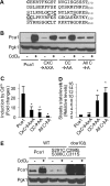
Roles for cysteine residues of Pca1p(250–350) in cadmium-induced Pca1p stabilization. A, amino acid sequence of Pca1p(251–350) with cysteine residues shown in bold. CXC and CC are underlined. Serine residues that exchange their positions with the first Cys residues of CXC and CC are underlined. B, the GPD1 gene promoter-mediated expression constructs of functional Pca1p with and without site-directed mutation of Cys residues in PCA1(250–350) were transformed to a pca1Δ strain. PCA1 was fused with N-terminal 3HA. Cells co-cultured with CdCl2 (1 μm for 30 min) at mid-log phase were subjected to total cell lysate preparation. Western blotting with anti-HA antibodies determined expression levels of Pca1p. Pgk1p levels were used to determine equal loading. A representative figure of four repeats is presented. C and D, quantification of the data presented in B. Cadmium-induced changes in expression levels of the indicated Pca1p were measured (C). Steady state levels of Pca1p in cells cultured without cadmium were presented as relative levels of control Pca1p (D). *, p < 0.01 compared with wild-type control Pca1p. E, expression levels of Pca1p possessing S291C, C298S, S306C, and C311S substitutions were determined in WT control and doa10Δ cells as described in B. Representative data of two independent experiments are presented.
FIGURE 4.
FIGURE 4.
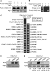
Identification of residues required for the functional role of Pca1p(250–350) as a degron. A, a GPD1 gene promoter-mediated expression construct of Yor1p with and without fusion of Pca1p(1–392) at the N terminus (Pca1(1–392)-Yor1) was transformed into BY4741 yeast cells. GFP was fused in-frame at the N terminus. Western blots using anti-GFP antibodies determined expression levels of Pca1p(1–392)-Yor1p in cells with and without cadmium (CuCl2) co-culture. B, growth of cells expressing empty vector and Pca1p(1–392)-Yor1p was examined on SC medium with and without supplementation of oligomycin and CdCl2 at the indicated concentrations. Cells at mid-log phase were spotted on solid medium, and cell growth was photographed in 2 days. C, PCA1(250–350) fragments containing random mutation(s) generated by error-prone PCR and flanked with HindIII and PstI sites were replaced with the corresponding fragment in PCA1(1–392)-YOR1. The plasmid library was transformed into yeast cells to select cells that can grow on solid medium containing a lethal concentration of oligomycin. Plasmids were retrieved from growing cells and subjected to sequencing. The growth of BY4741 yeast cells expressing empty vector, wild-type control Pca1p(1–392)-Yor1p, or selected plasmids containing amino acid substitution(s) within Pca1p(250–350) was examined on solid SC medium containing oligomycin (1 μg/ml). Cell growth was photographed in 3 days. The relative oligomycin resistance of cells was rated using cells expressing Pca1p(1–392)-Yor1p containing Pca1p(I299N) substitution as the highest (5, full growth) and wild-type control Pca1p(1–392)-Yor1p as the lowest (0, no growth). Several plasmids contained more than one mutation. The quantitation was confirmed by at least two independent experiments. D and E, Western blot using anti-HA antibodies determined steady-state expression levels of Pca1p with and without the indicated site-directed mutations. 3HA was fused at the N terminus (3HA-Pca1p). The GPD1 gene promoter-mediated expression constructs of 3HA-Pca1p were transformed into a pca1Δ strain. Cell lysates were subjected to Western blotting using anti-HA and -Pgk1p antibodies. A representative figure of four experiments is presented. E, the results of four experiments are quantitated and presented as -fold change in expression levels. *, p < 0.01.
FIGURE 5.
FIGURE 5.
Alignment of the Pca1p(271–320) sequence with the corresponding sequence of Pca1p-like proteins. Aspergillus fumigatus (A.f.), Gibberella zeae (G.z.), Penicillium janthinellum (P.j.), Aspergillus clavatus (A.c.), Botryotinia fuckeliana (B.f.), and Sclerotinia sclerotiorum (S.s.) possess Pca1p-like proteins that correspond to GenBank numbers 3510005, 2791550, 156536662, 4708560, 5432339, and 5485834, respectively. The amino acid sequence of Pca1p(271–320) was aligned with the corresponding sequence of those in the identified proteins. Asterisks indicate that all sequences aligned contain the same residue at that location. The conserved residues, including four Cys, are also highlighted in bold. A double dot (:) indicates conservation among the displayed sequences with a single exception. A single dot (.) represents a low level of conservation of amino acid type.
FIGURE 6.
FIGURE 6.
Prediction of the secondary structure of Pca1p(250–350) and amino acid substitution affecting Pca1p expression. A, the predicted structure of Pca1p(250–350). Seven Cys residues are indicated. B, distribution of amino acids within the Pca1p(271–306) helix predicted by helical wheel projection analysis. Filled diamonds, circles, triangles, and pentagons indicate hydrophobic, hydrophilic, potentially negative charged, and potentially positively charged residues, respectively. C, predicted secondary structure of Pca1p(271–320). The underlined residues indicate confirmed substitutions that affect Yor1p stabilization and Yor1p-mediated oligomycin resistance when Pca1p(1–350) containing the individually substitution is fused with Yor1p. D, perturbation of the secondary structure of Pca1p(271–320) by I299N substitution. E, formation of a hydrophobic patch by amino acids within Pca1p(271–306) obtained by hydrophobic cluster analysis. Amino acids that are involved in the formation of the hydrophobic patch are indicated by the outline. The amino acids are presented by their one-letter identification, except for glycine residues, which are represented by black diamonds, serines, which are black squares with a dot in the middle, and threonines, which are represented by black open squares. F, disruption of a hydrophobic patch within Pca1p(271–320) by I299N substitution.
FIGURE 7.
FIGURE 7.
Physical interaction between Pca1(250–350)p and a Hsp70p in a cadmium- and amino acid composition-dependent manner. A, yeast cells expressing Pca1p tagged with two c-myc epitopes (Pca1p-2Myc) and Ssa1p tagged with a triple HA epitope (3HA-Ssa1p) were co-cultured with and without CdCl2 (1 μm) for 1 h, and Pca1p(L296S)-2Myc (no CdCl2) was treated with the membrane-permeable cross-linker dimethyl 3,3′-dithiobispropionimidate (100 μg/ml). Cell lysates were subjected to Western blotting using anti-HA and anti-myc antibodies. Pgk1p was detected using specific antibodies to determine equal loading. Immunoprecipitation (IP) of Pca1p-2Myc in the lysates was carried out using anti-myc-conjugated beads (Pierce). Pca1p-2Myc and 3HA-Ssa1p in the eluted samples were detected by Western blotting. B, Western blotting analysis of Pca1p in WT and ssa1Δ cells with and without cadmium co-culture (1 μm CdCl2, 1 h). *, p < 0.05; **, p < 0.01; relative to WT control (first lane). C, Western blot of Pca1p expressed in the indicated yeast strains that were cultured at permissive (23 °C) and restrictive (37 °C) temperatures for 2 h. D, the indicated strains carrying an empty vector or Pca1p expression plasmid were co-cultured with cadmium (1 μm CdCl2, 30 min). Cellular cadmium levels were measured. *, p < 0.05; **, p < 0.01; relative to a corresponding strain containing an empty vector (−Pca1). #, p < 0.01 relative to the WT and ssa2,3,4Δ strain expressing Pca1p. A representative figure and/or mean ± S.D. of two (A) or three (B, C, and D) experiments are presented.

Similar articles

Cited by

References

    1. Ravid T., and Hochstrasser M. (2008) Diversity of degradation signals in the ubiquitin-proteasome system. Nat. Rev. Mol. Cell Biol. 9, 679–690 - PMC - PubMed
    1. Hochstrasser M. (1996) Protein degradation or regulation: Ub the judge. Cell 84, 813–815 - PubMed
    1. Zhang K., and Kaufman R. J. (2004) Signaling the unfolded protein response from the endoplasmic reticulum. J. Biol. Chem. 279, 25935–25938 - PubMed
    1. Babst M. (2014) Quality control: quality control at the plasma membrane: one mechanism does not fit all. J. Cell Biol. 205, 11–20 - PMC - PubMed
    1. Ansari N., and Khodagholi F. (2013) Molecular mechanism aspect of ER stress in Alzheimer's disease: current approaches and future strategies. Curr. Drug Targets 14, 114–122 - PubMed

Publication types

MeSH terms

LinkOut - more resources