. 2016 Jun 17;291(25):12991-3004.
doi: 10.1074/jbc.M116.721977. Epub 2016 Apr 8.

Structural Determinants of Binding the Seven-transmembrane Domain of the Glucagon-like Peptide-1 Receptor (GLP-1R)

Affiliations

Structural Determinants of Binding the Seven-transmembrane Domain of the Glucagon-like Peptide-1 Receptor (GLP-1R)

Dehua Yang et al. J Biol Chem. .

Abstract

The glucagon-like peptide-1 receptor (GLP-1R) belongs to the secretin-like (class B) family of G protein-coupled receptors. Members of the class B family are distinguished by their large extracellular domain, which works cooperatively with the canonical seven-transmembrane (7TM) helical domain to signal in response to binding of various peptide hormones. We have combined structure-based site-specific mutational studies with molecular dynamics simulations of a full-length model of GLP-1R bound to multiple peptide ligand variants. Despite the high sequence similarity between GLP-1R and its closest structural homologue, the glucagon receptor (GCGR), nearly half of the 62 stably expressed mutants affected GLP-1R in a different manner than the corresponding mutants in GCGR. The molecular dynamics simulations of wild-type and mutant GLP-1R·ligand complexes provided molecular insights into GLP-1R-specific recognition mechanisms for the N terminus of GLP-1 by residues in the 7TM pocket and explained how glucagon-mimicking GLP-1 mutants restored binding affinity for (GCGR-mimicking) GLP-1R mutants. Structural analysis of the simulations suggested that peptide ligand binding mode variations in the 7TM binding pocket are facilitated by movement of the extracellular domain relative to the 7TM bundle. These differences in binding modes may account for the pharmacological differences between GLP-1 peptide variants.

Keywords: G protein-coupled receptor (GPCR); glucagon; molecular dynamics; peptide interaction; site-directed mutagenesis.


Figures

FIGURE 1.
FIGURE 1.
Differential effects of human GLP-1R and human GCGR point mutations on GLP-1 and glucagon binding. A, comparison of the effects of 66 GLP-1R point mutations on GLP-1 binding and 66 GCGR mutations on glucagon binding, based on IC50 values in 125I-GLP-1 (for GLP-1R) and 125I-glucagon (for GCGR) displacement assays (including 50 GCGR mutants determined previously (11)). Mutated residues that had <4-fold change (blue), 4–10-fold increase (orange), and >10-fold increase (red) in IC50 values for GLP-1 binding to GLP-1R or glucagon binding to GCGR are shown. Mutant receptors with expression <30% of wild-type are colored gray. For each position, the results of mutations that show the most distinct differences between GLP-1R and GCGR are reported (see Table 1). The effects of previously reported human GLP-1R mutants are presented in supplemental Table S1 (7, 8, 19, 20, 25, 28–30, 32) and rat GLP-1R mutation data in supplemental Table S2 (26, 27). The most conserved residues in TM helices 1–7 of class B GPCRs (19) are boxed. B–D, representative 125I-GLP-1/GLP-1R (B and D) and 125I-glucagon/GCGR (C) displacement dose-response curves. Data are expressed as a percentage of specific 125I-GLP-1 or 125I-glucagon binding in the presence of 3.57 pm unlabeled peptide. Each point (±S.E.) represents the mean value of at least three independent experiments done in triplicate (IC50 data presented in Table 1 and supplemental Tables S1 and S3).
FIGURE 2.
FIGURE 2.
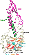
GLP-1R structural model explaining GLP-1R/GCGR-specific and ligand-dependent site-directed mutagenesis data. Mutations of homologous residues/positions in GLP-1R and GCGR but with differential effects on GLP-1 binding to GLP-1R versus glucagon binding to GCGR (Fig. 1A and supplemental Table S1) are mapped on a three-dimensional GLP-1R model and color-coded according their relative effects (ΔIC50) on GLP-1R versus GCGR binding as follows: >4-fold relative decrease (cyan), 4–10-fold relative increase (orange), and >10-fold relative increase (red) in IC50 values for GLP-1R mutant compared with the corresponding GCGR mutants. ΔIC50, the effect of a point mutant on binding of GLP-1 to GLP-1R or glucagon to GCGR, is defined as the ratio of IC50 values of wild-type and mutant. The relative effect of a mutation on ligand binding to GLP-1R versus GCGR is defined as the ratio of ΔIC50 (GLP-1R) and ΔIC50 (GLP-1R) values for the same residue position.
FIGURE 3.
FIGURE 3.
Probing the 7TM-binding site of GLP-1R with truncated peptide ligands. A, top view of effects of GLP-1R mutations on 125I-exendin-(9–39) displacement by GLP-1 (dark green), truncated GLP-1-(15–36), and exendin-4 (light green) (see Table 2) mapped on the three-dimensional GLP-1R model, color-coded according their effects (ΔIC50) on GLP-1R binding as follows: >4-fold decrease (cyan); 4-fold decrease to 4-fold increase (blue); 4–10-fold increase (orange); and >10-fold increase (red) in IC50 values for GLP-1R mutants. ΔIC50, the effect of a point mutant on binding of a specific peptide ligand to GLP-1, is defined as the ratio of IC50 values of wild-type and mutant. B, representative 125I-exendin-(9–39) displacement dose-response curves of GLP-1, exendin-4, and GLP-1-(15–36) at wild-type and mutant GLP-1R mutants used to derive IC50 data presented in Table 2. Data are expressed as a percentage of specific binding in the presence of 3.57 pm unlabeled peptide. Each point (±S.E.) represents the mean value of at least three independent experiments done in triplicate (IC50 data presented in Table 2 and supplemental Table S4).
FIGURE 4.
FIGURE 4.
Analysis of receptor-ligand interactions in GLP-1-bound GLP-1R and glucagon-bound GCGR. Comparison of 1000-ns MD simulations of GLP-1 (green) in GLP-1R (A–C) and glucagon (light green) in GCGR (D–F). Based on the analysis of receptor-ligand interaction patterns in GLP-1R (C) and GCGR (F), the structures of two representative snapshots at similar time frames (MD snapshots 1 and 2, indicated by green arrows and lines) of GLP-1R (A and B) and GCGR (D and E) MD simulations are shown. The analyses of H-bond interactions between positively and negatively charged groups (red), other H-bond interactions in which backbone groups (black) or only side-chain groups (gray) are involved, and apolar contacts (yellow) of specific residues in GLP-1/GLP-1R (C) and between specific residues in glucagon/GCGR (F) are shown.
FIGURE 5.
FIGURE 5.
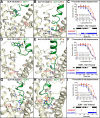
MD simulations explain combined GLP-1R and GLP-1 mutation studies. GLP-1 binding to GLP-1R mutants R1902.60bK (A), E3877.42bD (D), and Q2343.37bE (G) is restored by combination with glucagon-mimicking GLP-1 mutants Ala8Ser (B) and Glu9Gln (E and H), is determined by 125I-exendin-(9–39) displacement studies (C, F, and I), and is consistent with representative snapshots (A, B, D, E, G, and H) and interaction analyses (C, F, and I) of MD simulations studies (supplemental Fig. S3). Data in 125I-exendin-(9–39) displacement curves (C, F, and I) are expressed as a percentage of specific 125I-exendin-(9–39) binding in the presence of 3.57 pm unlabeled peptide. Each point (±S.E.) represents the mean value of at least three independent experiments done in triplicate (IC50 data presented in Table 3 and supplemental Table S5).
FIGURE 6.
FIGURE 6.
Alternative orientations of the ECD with respect to the 7TM domain accommodate different binding modes of glucagon-mimicking GLP-1 mutants in GCGR-mimicking GLP-1R mutants. Overlays of representative MD simulation snapshots of Ala8Ser GLP-1 mutant-bound E3877.42bD GLP-1R mutant (A), Glu9Gln GLP-1 mutant-bound R1907.42bK GLP-1R mutant (B), and Glu9Gln GLP-1 mutant-bound Q2343.37bE GLP-1R mutant with wild-type (WT) GLP-1-bound GLP-1R (C) are shown. The GLP-1 mutant (green) and ECD (magenta) and 7TM domain (light yellow) of the GLP-1R mutants are colored to allow comparison with the WT GLP-1 and GLP-1R (dark gray). The structures are superimposed onto the original model of the GLP-1R·GLP-1 complex using the main-chain atoms of residues I1461.41b–I1611.56b (TM1), F1842.53b–F1952.65b (TM2), L2313.34b–L2453.48b (TM3), S2714.47b–P2834.59b (TM4), L3115.41b–R3265.56b (TM5), K3516.40b–G3616.50b (TM6), and F7.48b–I4007.55b (TM7). The side views of the receptor·ligand complexes at the top of each panel are similar to the view presented in Figs. 2 and 3. The extracellular views of the receptor·ligand complexes presented in the middle of each panel are rotated vertically 180° clockwise and horizontally 90° counter-clockwise, compared with the top view, and are compatible with the XY plane shown at the bottom of each panel, which shows the average positions (and S.D. error bars) of the COM projections of the extracellular end of transmembrane helices (open squares), the ECD (R24–K130) (filled circles), and the helical part of the peptide ligands (S14–V33) (filled squares) on the XY plane in the last 500-ns simulations of different receptor-ligand systems.

References

    1. Meier J. J. (2012) GLP-1 receptor agonists for individualized treatment of type 2 diabetes mellitus. Nat. Rev. Endocrinol. 8, 728–742 - PubMed
    1. Moller D. E. (2001) New drug targets for type 2 diabetes and the metabolic syndrome. Nature 414, 821–827 - PubMed
    1. Parthier C., Reedtz-Runge S., Rudolph R., and Stubbs M. T. (2009) Passing the baton in class B GPCRs: peptide hormone activation via helix induction? Trends Biochem. Sci. 34, 303–310 - PubMed
    1. Donnelly D. (2012) The structure and function of the glucagon-like peptide-1 receptor and its ligands. Br. J. Pharmacol. 166, 27–41 - PMC - PubMed
    1. Hollenstein K., de Graaf C., Bortolato A., Wang M. W., Marshall F. H., and Stevens R. C. (2014) Insights into the structure of class B GPCRs. Trends Pharmacol. Sci. 35, 12–22 - PMC - PubMed

Publication types

MeSH terms

LinkOut - more resources