

Regulating the Rate of Molecular Self-Assembly for Targeting Cancer Cells
- PMID: 27062481
- PMCID: PMC4939907
- DOI: 10.1002/anie.201600753
Regulating the Rate of Molecular Self-Assembly for Targeting Cancer Cells
Abstract
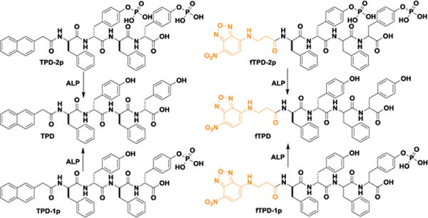
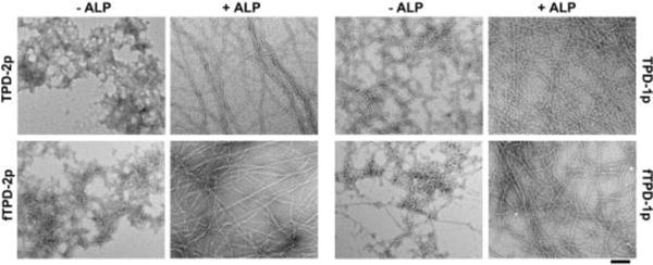
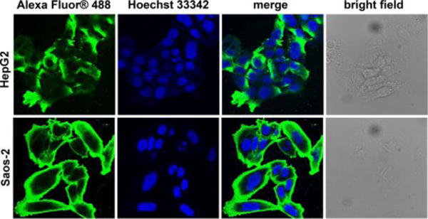
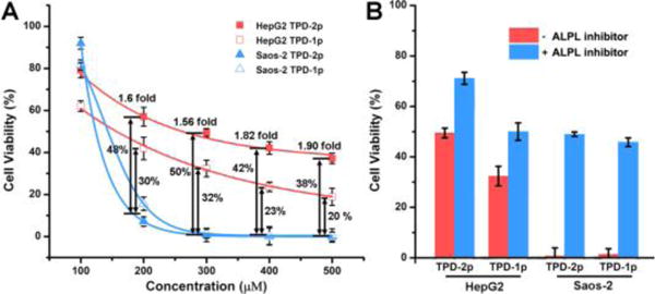
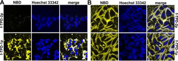
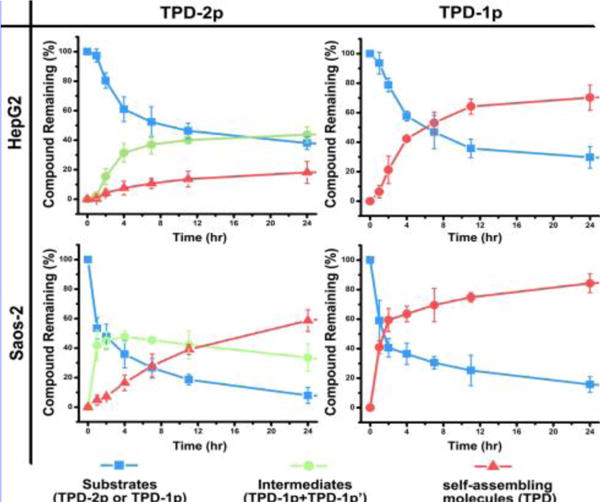
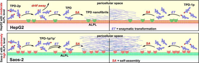
Besides tight and specific ligand-receptor interactions, the rate regulation of the formation of molecular assemblies is one of fundamental features of cells. But the latter receives little exploration for developing anticancer therapeutics. Here we show that a simple molecular design of the substrates of phosphatases-tailoring the number of phosphates on peptidic substrates-is able to regulate the rate of molecular self-assembly of the enzyme reaction product. Such a rate regulation allows selective inhibition of osteosarcoma cells over hepatocytes, which promises to target cancer cells in a specific organ. Moreover, our result reveals that the direct measurement of the rate of the self-assembly in a cell-based assay provides precise assessment of the cell targeting capability of self-assembly. This work, as the first report establishing rate regulation of a multiple-step process to inhibit cells selectively, illustrates a fundamentally new approach for controlling the fate of cells.
Keywords: cancer; enzymes; rate regulation; selective inhibition; self-assembly.
© 2016 WILEY-VCH Verlag GmbH & Co. KGaA, Weinheim.
Figures
References
-
- Yang ZM, Xu KM, Guo ZF, Guo ZH, Xu B. Adv Mater. 2007;19:3152–3156.
- Li J, Kuang Y, Shi J, Zhou J, Medina JE, Zhou R, Yuan D, Yang C, Wang H, Yang Z, Liu J, Dinulescu DM, Xu B. Angew Chem Int Edit. 2015;54:13307–13311. - PMC - PubMed
- Kuang Y, Shi J, Li J, Yuan D, Alberti KA, Xu Q, Xu B. Angew Chem Int Edit. 2014;53:8104–8107. - PMC - PubMed
- Shi J, Du X, Yuan D, Zhou J, Zhou N, Huang Y, Xu B. Biomacromolecules. 2014;15:3559–3568. - PMC - PubMed
- Zhou J, Du X, Li J, Yamagata N, Xu B. J Am Chem Soc. 2015;137:10040–10043. - PMC - PubMed
-
- Yang Z, Gu H, Fu D, Gao P, Lam JK, Xu B. Adv Mater. 2004;16:1440–1444.
- Toledano S, Williams RJ, Jayawarna V, Ulijn RV. J Am Chem Soc. 2006;128:1070–1071. - PubMed
- Yang Z, Liang G, Xu B. Acc Chem Res. 2008;41:315–326. - PubMed
- Miao X, Cao W, Zheng W, Wang J, Zhang X, Gao J, Yang C, Kong D, Xu H, Wang L, Yang Z. Angew Chem, Int Ed. 2013;52:7781–7785. - PubMed
- Zelzer M, Todd SJ, Hirst AR, McDonald TO, Ulijn RV. Biomater Sci. 2013;1:11–39. - PubMed
- Cheetham AG, Keith D, Zhang P, Lin R, Su H, Cui H. Curr Cancer Drug Targets. 2015 - PMC - PubMed
- Cui L, Tokarz D, Cisek R, Ng KK, Wang F, Chen J, Barzda V, Zheng G. Angew Chem, Int Ed. 2015;54:13928–13932. - PubMed
Publication types
MeSH terms
Substances
Grants and funding
LinkOut - more resources
Full Text Sources
Other Literature Sources
