. 2016 Apr 21;165(3):704-14.
doi: 10.1016/j.cell.2016.03.028. Epub 2016 Apr 7.

Mechanism of NMDA Receptor Inhibition and Activation

Affiliations

Mechanism of NMDA Receptor Inhibition and Activation

Shujia Zhu et al. Cell. .

Abstract

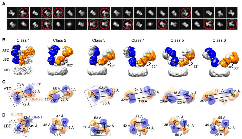
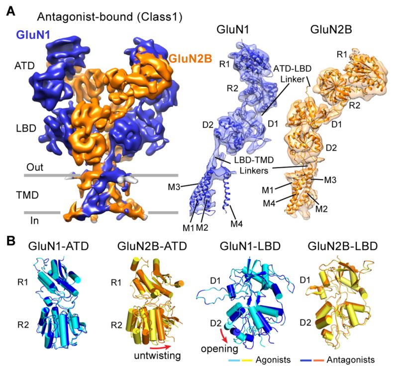
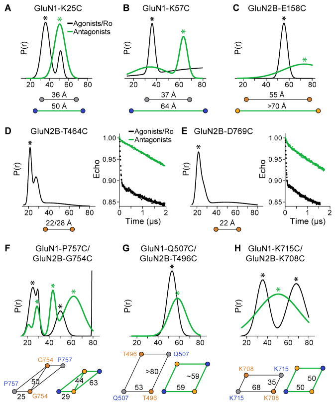
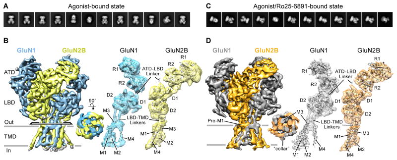
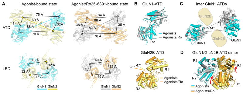
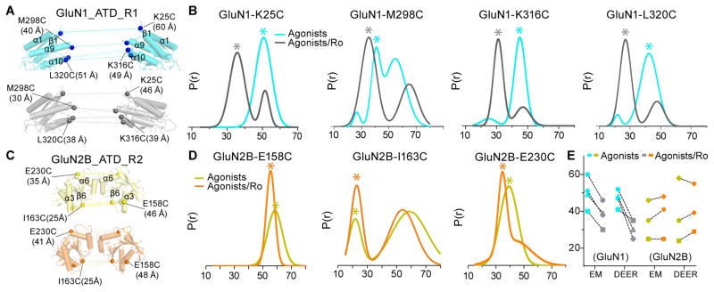
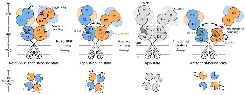
N-methyl-D-aspartate receptors (NMDARs) are glutamate-gated, calcium-permeable ion channels that mediate synaptic transmission and underpin learning and memory. NMDAR dysfunction is directly implicated in diseases ranging from seizure to ischemia. Despite its fundamental importance, little is known about how the NMDAR transitions between inactive and active states and how small molecules inhibit or activate ion channel gating. Here, we report electron cryo-microscopy structures of the GluN1-GluN2B NMDA receptor in an ensemble of competitive antagonist-bound states, an agonist-bound form, and a state bound with agonists and the allosteric inhibitor Ro25-6981. Together with double electron-electron resonance experiments, we show how competitive antagonists rupture the ligand binding domain (LBD) gating "ring," how agonists retain the ring in a dimer-of-dimers configuration, and how allosteric inhibitors, acting within the amino terminal domain, further stabilize the LBD layer. These studies illuminate how the LBD gating ring is fundamental to signal transduction and gating in NMDARs.


Figures

Figure 1
Figure 1. Cryo-EM Structures of Antagonist-Bound Receptors
(A) Representative 2D class average images of the DCKA/D-APV-bound state. Conformational heterogeneity with highly mobile extracellular domains are circled. (B) Six distinct classes of three-dimensional reconstruction density maps of DCKA/D-APV-bound GluN1-GluN2B (one subunit of each highlighted in blue and orange, respectively), showing antagonist-induced conformational changes of the ECDs. Rotation angles based on the inertial axis of mass weighting of the GluN2B-LBDs between the class 1 and the other five classes are indicated. (C–D) Measurements of the distances from the center of masses (shown as spheres) between GluN1 and GluN2B subunits are indicated by scale bars for the classes represented above. (C) The distances between the heterodimers in the ATD layer increase, while the distances within the dimer remain relatively unchanged. (D) The distance changes result in a transition from a twofold to a pseudo-fourfold symmetry on the LBD layer. The dimer units are highlighted by dashed box in class 1 and class 3. Views are ‘top down’ in (C) and (D).
Figure 2
Figure 2. Competitive Antagonist Binding Induces Clamshell Conformational Changes
(A) The 3D reconstruction density map of the DCKA/D-APV-bound GluN1-GluN2B receptor (in blue and orange, respectively), referring to class 1 in figure 1. View is perpendicular to the overall 2-fold axes of symmetry. Right panels are corresponding coordinate fits into the reconstruction map of the R1/R2 lobes of ATD, D1/D2 lobes of LBD, and TMD of the GluN1 or GluN2B subunits, independently. (B) Superimposition of the R1 lobes of the antagonist- and agonist-bound GluN1-ATD and GluN2B-ATD models (left panels), showing the untwisting motions of the GluN2B R2 lobe in the antagonist-bound state. Superimposition of the D1 lobes of the antagonist- and agonist-bound states of the GluN1-LBD and GluN2B-LBD models (right panels), showing the opening of the GluN1 D2 lobe in the DCKA-bound state.
Figure 3
Figure 3. Conformational Changes in the Antagonist-Bound State Monitored by DEER
(A–C) Probability distributions of DEER distances determined from DEER decays of MTSSL labeled antagonist- and agonist/Ro25-6981-bound states of GluN1-K25C (A), GluN1-K57C (B) and GluN2B-E158C (C) receptors. Distance changes for agonist/Ro25-6981 (in black) and antagonist (in green) are indicated. (D–E) DEER data of MTSSL-labeled GluN2B-T464C (D) and GluN2B-D769C (E). Peak-normalized echo decays and fits are shown on right. Measurements for agonist/Ro25-6981 are shown in black. (F–G) Probability distributions of DEER distances determined from DEER decays of MTSSL labeled antagonist- and agonist/Ro25-6981-bound states of GluN1-P757C/GluN2B-G754C (F), GluN1-Q507C/GluN2B-T496C (G), and GluN1-K715C/GluN2B-K708C (H) receptors. The asterisks indicate putative distances of the spin-labels. The diagrams below each distribution reflect the distances extracted from the peaks of DEER distribution.
Figure 4
Figure 4. Cryo-EM Structures of Agonist- and Agonist/Ro25-6981-Bound Receptors
(A and C) Representative 2D class average images of the agonist-bound (A) and agonist/Ro25-6981-bound (C) states. (B and D) The three-dimensional reconstruction density maps of the GluN1-GluN2B receptors in the agonist-bound (GluN1 in cyan and GluN2B in yellow, respectively) and agonist/Ro25-6981-bound states (GluN1 in grey and GluN2B in orange, respectively). Top-down views of the TMD are inserted in the middle panel. Right panels are corresponding coordinate fits into the reconstruction maps of the R1/R2 lobes of ATD, D1/D2 lobes of LBD, and TMD of the GluN1 or GluN2B subunits, independently. View is perpendicular to the overall 2-fold axes of symmetry.
Figure 5
Figure 5. Conformational Changes Effected by Allosteric Inhibitor
(A) Agonist- and agonist/Ro25-6981-bound GluN1-GluN2B receptor models derived from the density maps are shown in cyan/yellow and grey/orange, respectively. Distances from the center of mass (spheres) between subunits for the ATD (top panels) and LBD (bottom panels) are indicated by scale bars. (B) Superimposition of the R1 lobes of the agonist- and agonist/Ro25-6981-bound states of the GluN1-ATD (top panel) and GluN2B-ATD (bottom panel) domains, showing the 20° opening (measured by dihedral angles connecting the Cα atoms of E101, Q147, E230 and Y277) of the GluN2B clamshell in the agonist-bound state. (C) Superimposition of one GluN1 for the agonist- and agonist/Ro25-6981-bound states, showing the intersubunit rotation of the other GluN1 clamshell-liked domain.(D) Superimposition of the GluN1-ATD of the agonist- and agonist/Ro25-6981-bound states within the ATD heterodimer, showing relative movement of the R1 and R2 lobes of GluN2B ATD.
Figure 6
Figure 6. Separation of GluN1 ATDs, but not GluN2 ATDs Detected by DEER
(A and C) Corresponding models from coordinate fits into the three-dimensional EM reconstruction ECD maps with side views of the R1 lobes of inter-GluN1 ATDs (A) and R2 lobes of inter-GluN2B ATDs (C) in complex with agonist (cyan in A; yellow in C) and agonist/Ro25-6981 (grey in A; orange in C). Distance between Cα atoms for GluN1 (Lys 25, Met 298, Lys 316 and Leu 320) and GluN2B (Glu 158, Ile 163 and Glu 230) are indicated. (B and D) Probability distributions of DEER distances determined from DEER decays of MTSSL labeled agonist- and agonist/Ro25-6981-bound GluN1-GluN2B of indicated receptor constructs. Asterisk indicates putative distances from the spin-labels under the given condition. (E) Summary of the distance changes in the agonist- and agonist/Ro25-6981-bound GluN1-GluN2B receptor states observed from the cryo-EM structures and DEER measurements.
Figure 7
Figure 7. Schematic Summary of ECD Conformational Changes in NMDAR
Shown are the conformational changes of the extracellular ATD and LBD layers associated with the transition from the apo state to the antagonist-bound state (right), the agonist-bound state (middle) and the agonist/Ro25-6981-bound (left). The bottom panel shows top-down views of various LBD conformations at different states. At present there is no structure for an apo-state and thus the schematic is grey.

References

    1. Armstrong N, Jasti J, Beich-Frandsen M, Gouaux E. Measurement of conformational changes accompanying desensitization in an ionotropic glutamate receptor. Cell. 2006;127:85–97. - PubMed
    1. Attwell D, Gibb A. Neuroenergetics and the kinetic design of excitatory synapses. Nature reviews Neuroscience. 2005;6:841–849. - PubMed
    1. Baron BM, Siegel BW, Slone AL, Harrison BL, Palfreyman MG, Hurt SD. [3H]5,7-dichlorokynurenic acid, a novel radioligand labels NMDA receptor-associated glycine binding sites. European journal of pharmacology. 1991;206:149–154. - PubMed
    1. Biasini M, Bienert S, Waterhouse A, Arnold K, Studer G, Schmidt T, Kiefer F, Cassarino TG, Bertoni M, Bordoli L, et al. SWISS-MODEL: modelling protein tertiary and quaternary structure using evolutionary information. Nucleic acids research. 2014;42:W252–258. - PMC - PubMed
    1. Chaudhry C, Weston MC, Schuck P, Rosenmund C, Mayer ML. Stability of ligand-binding domain dimer assembly controls kainate receptor desensitization. The EMBO journal. 2009;28:1518–1530. - PMC - PubMed

Publication types

MeSH terms