. 2016 Apr 11:7:11242.
doi: 10.1038/ncomms11242.

The Ku-binding motif is a conserved module for recruitment and stimulation of non-homologous end-joining proteins

Affiliations

The Ku-binding motif is a conserved module for recruitment and stimulation of non-homologous end-joining proteins

Gabrielle J Grundy et al. Nat Commun. .

Abstract

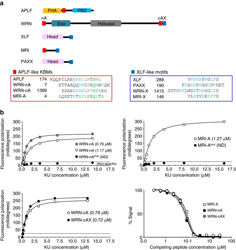
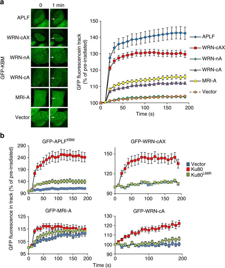
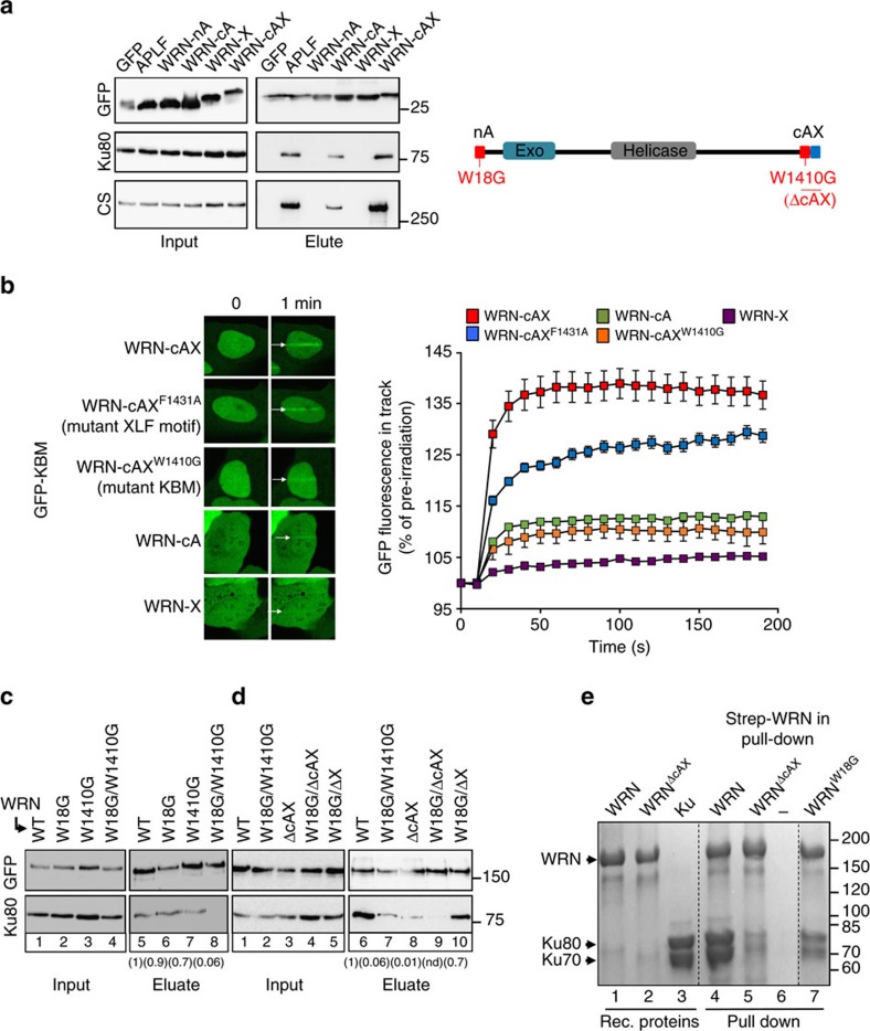
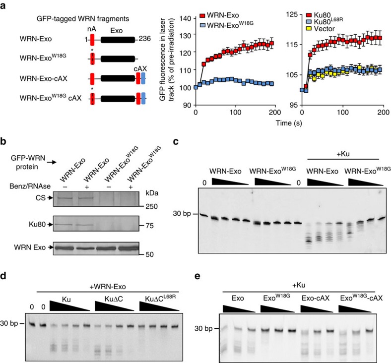
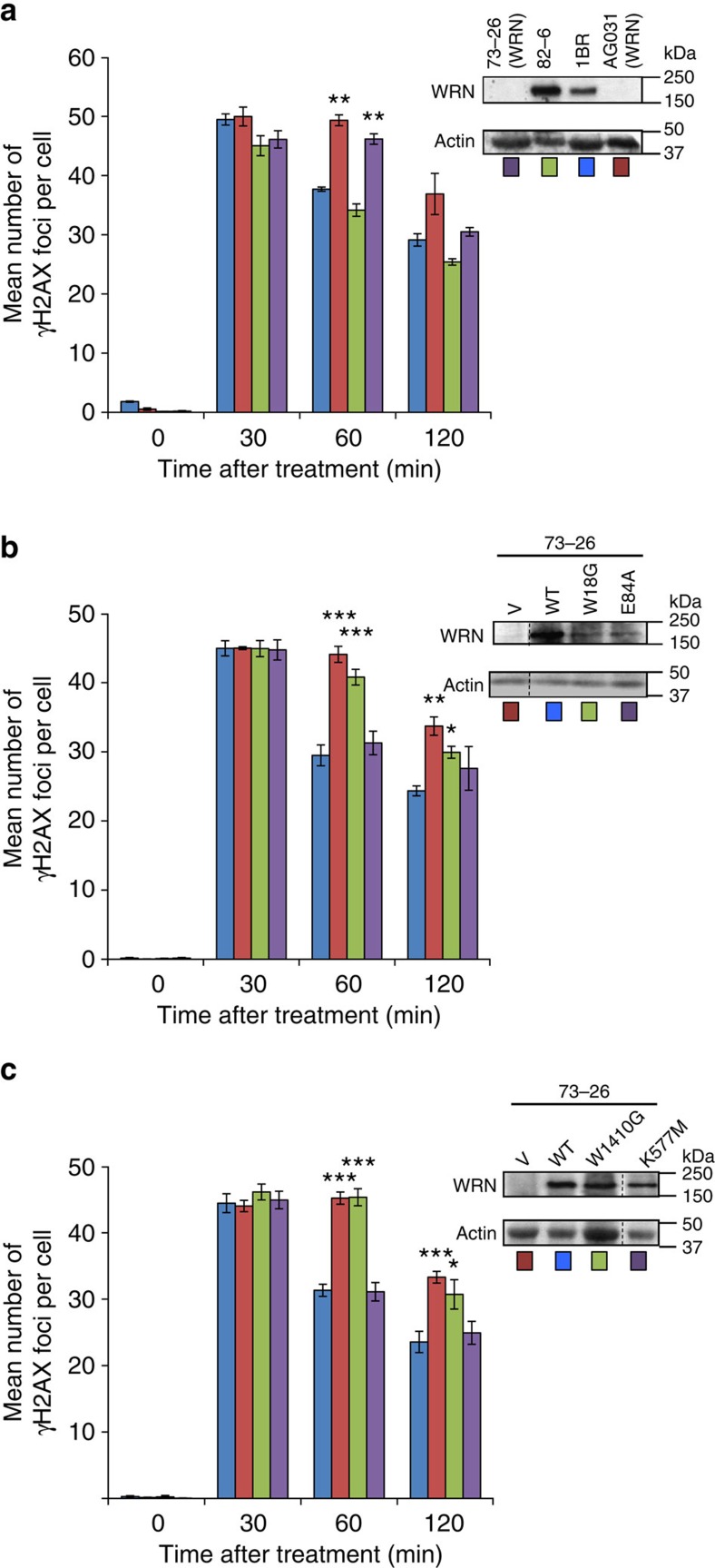
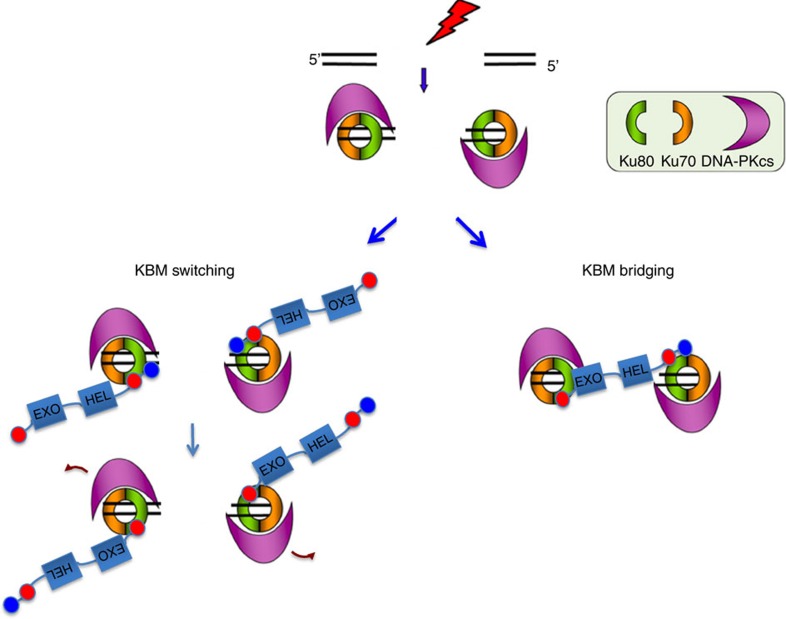
The Ku-binding motif (KBM) is a short peptide module first identified in APLF that we now show is also present in Werner syndrome protein (WRN) and in Modulator of retrovirus infection homologue (MRI). We also identify a related but functionally distinct motif in XLF, WRN, MRI and PAXX, which we denote the XLF-like motif. We show that WRN possesses two KBMs; one at the N terminus next to the exonuclease domain and one at the C terminus next to an XLF-like motif. We reveal that the WRN C-terminal KBM and XLF-like motif function cooperatively to bind Ku complexes and that the N-terminal KBM mediates Ku-dependent stimulation of WRN exonuclease activity. We also show that WRN accelerates DSB repair by a mechanism requiring both KBMs, demonstrating the importance of WRN interaction with Ku. These data define a conserved family of KBMs that function as molecular tethers to recruit and/or stimulate enzymes during NHEJ.


Figures

Figure 1
Figure 1. Conserved Ku-binding motifs (KBMs).
(a) Cartoon of NHEJ proteins containing putative APLF-like KBMs (red squares) and/or the related XLF-like motif (blue squares). Peptide sequences (lower panels) highlight the conserved basic (blue), hydrophobic (green), proline (purple), and tryptophan/phenylalanine (green bold) residues characteristic of these motifs. The tryptophan residues mutated for the fluorescence polarization (FP) assays described below are underlined. (b) Top and bottom left panels, FP assays measuring direct interaction between synthetic fluorescein-labeled peptides (100 nM) encoding the indicated KBMs and the indicated concentration of Ku heterodimer (KuΔC). Peptide sequences are those shown in a, but additionally preceded at the N terminus by fluorescein-GGYG. Mutant peptides have alanine instead of tryptophan at the positions underlined in a. WRN-cAX peptide encodes both the APLF-like KBM (residues 1,399–1,414) and XLF-like motif (residues 1,415–1,432) from the WRN C terminus. Bottom right panel, MRI-A, WRN-nA or WRN-cAX peptides (2.1 μM) were employed in FP competition assays with KuΔC (1 μM) and the indicated concentration (X-axis) of unlabeled APLF KBM peptide. All data points are the mean of three independent experiments (±1 s.d.). Kd values are indicated in parentheses (±1 s.d.) unless too weak to be determined (‘ND').
Figure 2
Figure 2. KBM accumulation at sites of UVA laser-induced chromosome damage.
(a) U2-OS cells were transiently transfected with expression constructs encoding GFP alone (Vector) or the indicated GFP-tagged KBM and subjected to UVA laser-induced micro-irradiation. The expressed peptide sequences for each KBM were APLF (177–193), WRN-cAX (1,399–1,432), WRN-nA (10–23), WRN-cA (1,399–1,414), MRI-A (6–19). Images were captured immediately before and at 10 s intervals following treatment. Representative images are shown on the left and quantified data on the right. (b) Ku80−/− mouse embryonic fibroblasts (MEFs) were co-transfected with expression constructs encoding the GFP-tagged KBM from APLF or the indicated GFP-tagged APLF-like KBMs from WRN or MRI, mRFP-Ku70, and either mRFP (‘vector'), mRFP-Ku80 or mRFP-Ku80L68R. Cells were micro-irradiated with UVA as above. All data are the mean GFP fluorescence (±s.e.m.) in the laser track relative to the mean GFP fluorescence before irradiation (set at 100%) from >20 cells per experiment.
Figure 3
Figure 3. The WRN C-terminal KBM and XLF-like motif bind Ku protein complexes cooperatively.
(a) HEK293T cells were co-transfected with expression constructs encoding GFP or the indicated GFP-tagged KBMs and GFP-tagged proteins recovered using GFP-TRAP beads. Aliquots of the input and eluate samples were fractionated by SDS–PAGE and immunoblotted for GFP, Ku80 and DNA–PKcs (CS). Right, cartoon depicting WRN and the position of the KBMs and XLF-like motif and the mutations employed in these experiments. (b) HEK293T cells were transfected with expression constructs encoding the indicated wild-type or mutated GFP-tagged WRN C-terminal KBM, XLF-like motif (‘X'), or KBM plus XLF-like motif in tandem. Cells were micro-irradiated with UVA as in Fig. 2. Representative images (left) and quantification (right) are shown. All quantified data are the mean GFP fluorescence (±s.e.m.) in the laser track relative to the mean GFP fluorescence before irradiation (set at 100%) from >20 cells per experiment. (c,d) Expression constructs encoding full-length wild-type (‘WT') GFP–WRN or derivatives harbouring the indicated point mutations in the N-terminal KBM (W18G), C-terminal KBM (W1410G) or deleted C-terminal tandem domain (ΔcAX) or XLF-like motif (ΔX ) were transfected into HEK293T cells and recovered using GFP-TRAP beads. Input and eluates were immunoblotted for GFP and Ku80. Numbers in parentheses are the fraction of Ku co-precipitated by the indicated GFP-tagged WRN protein, relative to wild-type WRN, quantified by ImageJ. Data are from two to six independent experiments, except for W18G/ΔcAX in which Ku recovery was too low to be determined (‘nd'). (e) Direct interaction of purified full-length Strep-tagged WRN with recombinant human Ku. Recombinant Strep-tagged WRN, WRNΔcAX or WRNW18G was immobilized on Streptavidin Mag sepharose beads and incubated with recombinant Ku heterodimer. Aliquots of the recombinant proteins employed in the experiment are shown on the left (lanes 1–3) and proteins pulled down by the indicated Strep-tagged WRN protein are shown on the right (lanes 4–7). Lane 6 contains the proteins recovered in a control pull-down that lacked Strep-tagged WRN. Proteins were fractioned by SDS–PAGE and stained with Coomassie Blue.
Figure 4
Figure 4. The WRN N-terminal KBM promotes WRN exonuclease activity.
(a) Left, cartoon illustrating the GFP-tagged truncated recombinant WRN proteins employed in these experiments. The WRN N-terminal (‘nA') and C-terminal (‘cA') KBMs are indicated by red boxes and XLF-like motif (‘X') by a blue box. The exonuclease domain is indicated by a black box, and the position of the KBM mutation (W18G) by an asterisk and dotted line. Middle, U2-OS cells transiently expressing the indicated recombinant GFP-tagged WRN protein were imaged for GFP before and after UVA microirradiation, as in Fig. 2. Right, Ku80−/− MEFs transiently co-expressing GFP-tagged WRN-Exo, RFP-Ku70, and either RFP (vector), RFP-Ku80 or RFP-Ku80L68R as indicated were micro-irradiated as in Fig. 1. Data are the mean GFP fluorescence (±s.e.m.) in the laser track relative to the mean GFP fluorescence before irradiation (set at 100%) from >20 cells per experiment. (b) The indicated GFP-tagged WRN proteins were recovered from transiently transfected HEK293T cell lysates pre-treated or not as indicated with Benzonase and RNAse in pull-down assays using GFP-TRAP beads. Aliquots of the bead eluate were fractionated by SDS-PAGE and silver stained to detect GFP-WRN, GFP-WRNW18G, Ku80, and DNA-PKcs (‘CS'). (c) Cy3-labeled 30 bp duplex oligonucleotide (20 nM) with a 5′ overhang was incubated with 500, 100, 20 or 5 nM HIs-tagged WRN-Exo or WRN-ExoW18G in the absence or presence of 100 nM Ku heterodimer (Ku70/Ku80, ‘Ku') and 5 mM MgCl2. Exonuclease products were resolved on a 16% TBE-Urea gel. (d) Exonuclease assays were conducted as above in the presence of 5 mM MgCl2 using 10 nM His-tagged WRN-Exo and 100, 20, 4 or 0.8 nM of either Ku heterodimer (Ku70/Ku80; ‘Ku'), KuΔC heterodimer (Ku70/Ku80ΔC; ‘KuΔC'), or mutant KuΔC heterodimer harbouring the Ku80 mutation, L68R (KuΔCL68R). (e) Exonuclease assays were conducted as above using 100, 20 and 4 nM of the indicated His-tagged WRN protein and 10 nM wild-type Ku heterodimer (Ku70/Ku80; ‘Ku') in 5 mM Mg2+.
Figure 5
Figure 5. WRN KBMs accelerate DSB repair.
(a) Confluence-arrested (G0/G1) hTERT-immortalised fibroblasts from two WRN patients (73–26 and AG03141) and normal controls (82–6 and 1BR) were treated with γ-rays (2 Gy) and γH2Ax foci counted at the time-points indicated. Inset, Actin and WRN protein levels in the indicated cell lines. (b) WRN cells (73–26) stably transduced with empty vector (V) or vector encoding wild-type (WT) WRN, WRNW18G harbouring a mutated N-terminal KBM (W18G), or WRN harbouring a mutated exonuclease domain WRNE84A (E84A), were examined for DSB repair rates as described above. (c) Werner syndrome cells (73–26) stably transduced with empty vector (V) or vector encoding WT WRN, WRNW1410G harbouring a mutated C-terminal KBM (W1410G), or WRN harbouring a mutated helicase domain (WRNK577M) were examined as above. Data points are the mean (±s.e.m.) number of foci per cell from four independent experiments. *P<0.05, **P<0.01, ***P<0.001 by paired t-test when compared with WT cells.
Figure 6
Figure 6. A Model for WRN KBM function during NHEJ.
Top, DNA–PK holoenzyme binds to a DSB. Bottom left, WRN is recruited into DNA–PK complexes by high affinity interaction between the C-terminal KBM (red circle) and the hydrophobic pocket in the vWA domain of Ku80. The XLF-like motif (blue circle) functions cooperatively, perhaps stabilizing the association of Ku with DNA–PKcs. Following autophosphorylation, DNA–PKcs dissociates and the C-terminal KBM is replaced by the N-terminal KBM to stimulate WRN 3′-exonucease activity. Bottom right, The N-terminal and C-terminal KBMs bind two Ku molecules simultaneously, bridging the DSB. Note that WRN may fulfil both enzymatic and structural roles during NHEJ.

References

    1. Cannan W. J. & Pederson D. S. Mechanisms and consequences of double-strand DNA break formation in chromatin. J. Cell. Physiol. 231, 3–14 (2016). - PMC - PubMed
    1. Soulas-Sprauel P. et al. V(D)J and immunoglobulin class switch recombinations: a paradigm to study the regulation of DNA end-joining. Oncogene 26, 7780–7791 (2007). - PubMed
    1. Bunting S. F. & Nussenzweig A. End-joining, translocations and cancer. Nat. Rev. Cancer 13, 443–454 (2013). - PMC - PubMed
    1. McKinnon P. J. & Caldecott K. W. DNA strand break repair and human genetic disease. Annu. Rev. Genomics Hum. Genet. 8, 37–55 (2007). - PubMed
    1. Woodbine L., Gennery A. R. & Jeggo P. A. The clinical impact of deficiency in DNA non-homologous end-joining. DNA Repair (Amst) 16, 84–96 (2014). - PubMed

Publication types