. 2016 Jun;37(6):763-71.
doi: 10.1038/aps.2016.14. Epub 2016 Apr 11.

Methylophiopogonanone A suppresses ischemia/reperfusion-induced myocardial apoptosis in mice via activating PI3K/Akt/eNOS signaling pathway

Affiliations

Methylophiopogonanone A suppresses ischemia/reperfusion-induced myocardial apoptosis in mice via activating PI3K/Akt/eNOS signaling pathway

Fei He et al. Acta Pharmacol Sin. 2016 Jun.

Abstract

Aim: The dried tuber root of Ophiopogon japonicus has been used in the traditional Chinese medicine for treatment of myocardial ischemia and thrombosis. In this study we investigated the effects of methylophiopogonanone A (MO-A), a major homoisoflavonoid in Ophiopogon japonicus, on myocardial ischemia/reperfusion (I/R) injury.

Methods: Mice were pretreated with MO-A (10 mg·kg(-1)·d(-1), po) for 2 weeks and then subjected to transient occlusion of the left anterior descending coronary artery. Cardiac function was evaluated, and the infarct size and apoptosis index were assessed. The mechanisms underlying the cardio-protection of MO-A were analyzed in H9C2 rat cardiomyocytes subjected to hypoxia/reoxygenation (H/R). The cell viability and apoptosis were evaluated; apoptotic and relevant signaling proteins were analyzed. NO levels in the culture medium were assessed.

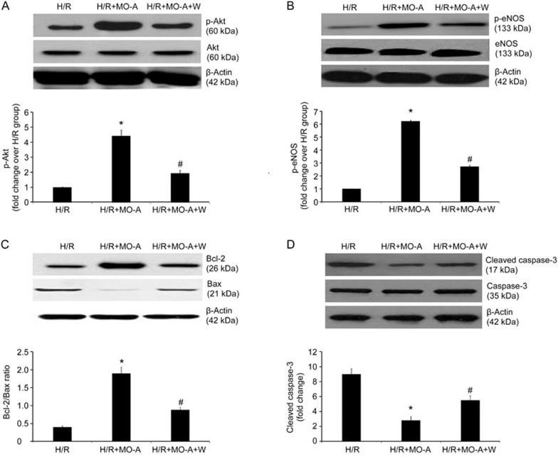
Results: In I/R mice, pretreatment with MO-A significantly reduced the infarct size (by 60.7%) and myocardial apoptosis (by 56.8%), and improved cardiac function. In H9C2 cells subjected to H/R, pretreatment with MO-A (10 μmol/L) significantly decreased apoptosis and cleaved caspase-3 expression, elevated the Bcl-2/Bax ratio and restored NO production. Furthermore, pretreatment with MO-A markedly increased the activation of PI3K/Akt/eNOS pathway in H9C2 cells subjected to H/R, and the protective effects of MO-A were abolished in the presence of the PI3K inhibitor wortmannin (100 nmol/L).

Conclusion: MO-A attenuates I/R-induced myocardial apoptosis in mice via activating the PI3K/Akt/eNOS signaling pathway.


Figures

Figure 1
Figure 1
MO-A reduced infarct size and apoptosis following myocardial I/R in mice. (A) Representative photomicrographs of TTC/Evan blue staining in heart tissues from mice in sham, I/R, and I/R+MO-A pretreatment group. (B) Representative micrographs of left mid-ventricular sections with TUNEL staining (arrows indicate TUNEL-positive nuclei). (C) MO-A reduced infarct size following myocardial I/R in mice. (D)MO-A inhibited apoptosis following myocardial I/R in mice. Values presented are mean±SEM. n=10 in each group. *P<0.05 vs sham operation. #P<0.05 vs I/R group.
Figure 2
Figure 2
MO-A preserved left ventricular function following myocardial I/R in mice. (A) Representative photographs of ultrasound cardiographs from the sham operation group and from mice that underwent I/R with or without MO-A pretreatment. (B–F) Cardiac function was measured by echocardiography or hemodynamic measurements before and after I/R. Values presented are mean±SEM. n=10 in each group. *P<0.05 vs sham operation. #P<0.05 vs I/R group. Left ventricular ejection fraction: EF; Left ventricular fraction shortening: FS; Maximal velocity of left ventricular pressure development: +dp/dt; Left ventricular end-diastolic pressure: LVEDP; Left ventricular systolic pressure: LVSP.
Figure 3
Figure 3
Assessment of apoptotic cell death and cell viability in cultured H9C2 cells. (A) Flow cytometry revealed that MO-A alleviated H/R-induced apoptosis of H9C2. The lower right (LR) region shows the early apoptotic cells (FITC+/PI-). (B) MO-A decreased H/R-induced apoptosis of H9C2 cells. (C) MO-A increased cell viability after H/R treatment. Values presented are mean±SEM. n=6 in each group. *P<0.05 vs sham operation. #P<0.05 vs H/R group.
Figure 4
Figure 4
MO-A activated the PI3K-Akt-eNOS pathway in H9C2 cells exposed to H/R. (A) Ratio of PI3K/β-actin. (B) Ratio of phosphorylated Akt/total Akt; and (C) ratio of phosphorylated eNOS/total eNOS. (D–F) p-Akt, p-eNOS and PI3K levels were normalized by Akt, eNOS and β-actin, respectively, and expressed as fold of the H/R group. Values presented are mean±SEM. n=6 in each group. *P<0.05 vs sham operation. #P<0.05 vs H/R group.
Figure 5
Figure 5
MO-A restored NO production, increased Bcl-2/Bax ratio and decreased cleaved caspase-3 in H9C2 cells exposed to H/R. (A) MO-A restored NO production in H9C2 cells exposed to H/R. (B) The protein levels of Bcl-2 and Bax and the ratio of Bcl-2/Bax. (C) The protein levels of cleaved caspase-3 and the ratio of cleaved caspase-3/β-actin. (D–E) Quantitative analysis of Bcl-2/Bax and cleaved caspase 3. Values presented are mean±SEM. n=6 in each group. *P<0.05 vs sham operation. #P<0.05 vs H/R group.
Figure 6
Figure 6
Effects of MO-A on apoptosis, cell viability and NO production were blocked by the PI3K/Akt inhibitor wortmannin. (A)Apoptosis rate of H9C2 cells after H/R determined by flow cytometry. (B)MO-A significantly reduced the apoptosis rate of H9C2 cells after H/R. (C) The anti-apoptotic effect of MO-A was blocked by co-treatment with the PI3K/Akt inhibitor wortmannin. (D–E) The effect of MO-A on cell viability was blocked by the PI3K/Akt inhibitor wortmannin. (F) The effect of MO-A on NO production was blocked by the PI3K/Akt inhibitor wortmannin. Values presented are mean±SEM. n=6 in each group. *P<0.05 vs H/R group. #P<0.05 vs H/R+MO-A group.
Figure 7
Figure 7
MO-A increased the Bcl-2/Bax ratio and decreased caspase-3 expression by activating the PI3K-Akt signaling pathway. (A–B) The expression levels of phosphorylated Akt and eNOS were reduced after H/R in H9C2 cells when cells were co-treated with the PI3K/Akt inhibitor wortmannin. (C) The protein levels of Bcl-2 and Bax and the ratio of Bcl-2/Bax. (D) The protein levels of cleaved caspase-3 and the ratio of cleaved caspase-3/β-actin. Values presented are mean±SEM. n=6 in each group. *P<0.05 vs H/R group. #P<0.05 vs H/R+MO-A group.

References

    1. Ibanez B, Heusch G, Ovize M, Van de Werf F. Evolving therapies for myocardial ischemia/reperfusion injury. J Am Coll Cardiol 2015; 65: 1454–71. - PubMed
    1. Yellon DM, Hausenloy DJ. Mechanisms of disease: myocardial reperfusion injury. New Engl J Med 2007; 357: 1121–35. - PubMed
    1. Heusch G. Molecular basis of cardioprotection: signal transduction in ischemic pre-, post-, and remote conditioning. Circ Res 2015; 116: 674–99. - PubMed
    1. Hernandez-Resendiz S, Palma-Flores C, De Los Santos S, Roman-Anguiano NG, Flores M, de la Pena A, et al. Reduction of no-reflow and reperfusion injury with the synthetic 17beta-aminoestrogen compound Prolame is associated with PI3K/Akt/eNOS signaling cascade. Basic Res Cardiol 2015; 110: 1. - PubMed
    1. Ma CJ, Li G, Zhang J, Zheng QS, Fan X, Wang ZH. An efficient combination of supercritical fluid extraction and high-speed counter-current chromatography to extract and purify homoisoflavonoids from Ophiopogon japonicus (Thunb) Ker-Gawler. J Sep Sci 2009; 32: 1949–56. - PubMed

MeSH terms