Review
. 2016 Jul:163:94-108.
doi: 10.1016/j.pharmthera.2016.03.013. Epub 2016 Apr 8.

Three-dimensional culture systems in cancer research: Focus on tumor spheroid model

Affiliations
Review

Three-dimensional culture systems in cancer research: Focus on tumor spheroid model

Sritama Nath et al. Pharmacol Ther. 2016 Jul.

Abstract

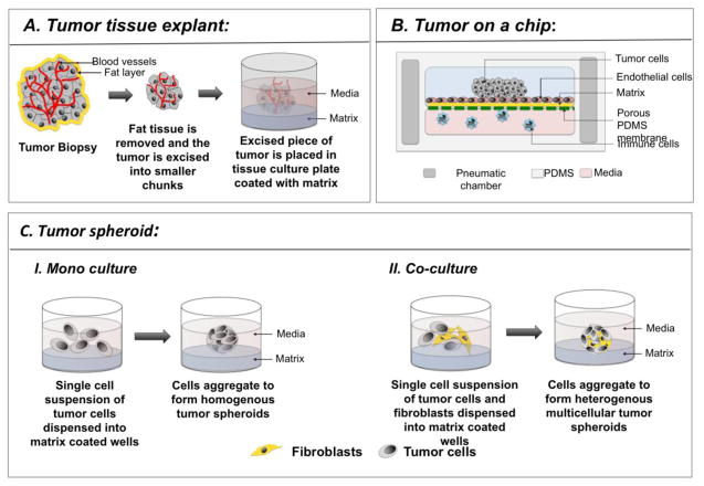
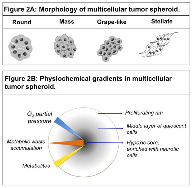
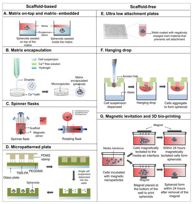
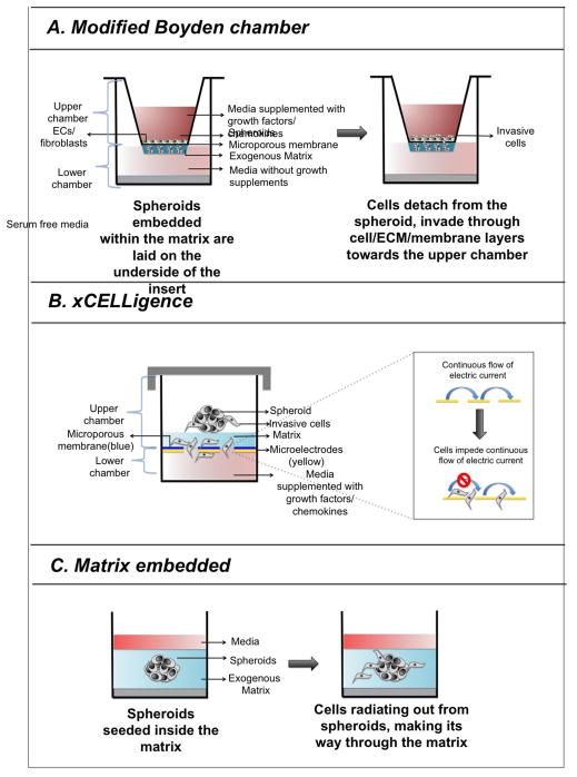
Cancer cells propagated in three-dimensional (3D) culture systems exhibit physiologically relevant cell-cell and cell-matrix interactions, gene expression and signaling pathway profiles, heterogeneity and structural complexity that reflect in vivo tumors. In recent years, development of various 3D models has improved the study of host-tumor interaction and use of high-throughput screening platforms for anti-cancer drug discovery and development. This review attempts to summarize the various 3D culture systems, with an emphasis on the most well characterized and widely applied model - multicellular tumor spheroids. This review also highlights the various techniques to generate tumor spheroids, methods to characterize them, and its applicability in cancer research.

Keywords: Apoptosis; High-throughput screening; Inflammatory breast cancer; Invasion; Oxidative stress; Tumor emboli.


Conflict of interest statement

Conflict of interest:

The authors have no conflict of interest.

Figures

Figure 1
Figure 1
Schematic representing the various 3D models of cancer. A. Excised tumor biopsy is processed to remove the excess fat and necrotic cells, and cut into small pieces. After washing the tumor in PBS, it is placed on a tissue culture plate that has been coated with a matrix, such as Matrigel of methylcellulose, to which the tumor sits atop firmly or is embedded. Media is added and the tumor is cultured for the duration of the experiment. B. “Tumor on a chip” represents a vasculature mimicking microfluidic device consisting of PDMS chambers with highly organized microchannels and pneumatic chamber (dark grey) on either sides. The microchannels (pink) contain media, in which immune cells and circulating tumor cells navigate. The top chamber contains matrix coated (yellow) porous membrane (green), with a monolayer of endothelial cells on top. The tumor cells are loaded through an inlet into the top chamber. Cells that have been genetically modified to express fluorescent protein can be observed in real time to monitor their functional changes, such as invasion, and migration. C. Schematic depicting tumor spheroid formation where tumor spheroids have been generated by culturing tumor cells alone or in combination with fibroblasts.
Figure 2
Figure 2
A: Schematic representing the various morphologies of tumor spheroids. Tumor spheroids adapt various shapes depending upon the culture conditions and inherent nature of the tumor cells. B. Schematic representing the presence of physiochemical gradients in a spheroid and the resulting complexity in spheroid composition. Availability of the O2 (blue triangle) and nutrients (yellow triangle) diminishes with increasing depth of the spheroid. Whereas, metabolic waste accumulation (red triangle) is highest at the core compared to the peripheral layer of the spheroid. Hypoxia at the core (blue arrow) of the spheroid triggers necrosis (black circle), which precedes a layer of apoptotic cells. A middle layer of quiescent cells is sandwiched between the necrotic core and the peripheral layer of proliferating cells.
Figure 3
Figure 3
Schematic explains the various methods to generate tumor spheroids. Figures AD represent scaffold-based methods, whereas figures E–F represent scaffold-free methods.
Figure 4
Figure 4. Schematic representing the various spheroid-based invasion assays
A. In modified-Boyden chamber based invasion assay, a thin layer of spheroid and matrix mixture is coated on the. Within 72 hours, cells begin to disseminate from the spheroid, proteolytically cleave through the matrix and migrate towards the bottom chamber through a matrix coated porous membrane located at the interface of upper and lower chambers. A layer of endothelial cells placed on top of the matrix coated porous membrane allow to determine the invasive potential of the cells through matrix and cellular barriers. B. The schematic represents a CIM plate, which has similar components as Boyden chamber. However, the detection system is different in xCelligence, where invasive potential of tumor cells is determined by measuring electrical impendence imposed by them. C. Spheroid is seeded on a layer of matrix, such as Matrigel, Methylcellulose, and Collagen type I, which is followed by a second layer of matrix. Cells from the embedded spheroid detach from the spheroid, radiating outward, which is measured in real time by Celigo Cytometer.

References

    1. Abuelba H, Cotrutz CE, Stoica BA, Stoica L, Olinici D, Petreus T. In vitro evaluation of curcumin effects on breast adenocarcinoma 2D and 3D cell cultures. Romanian Journal of Morphology & Embryology. 2015;56:71–76. - PubMed
    1. Ahmed EM. Hydrogel: Preparation, characterization, and applications: A review. Journal of Advanced Research. 2015;6:105–121. - PMC - PubMed
    1. Albanese A, Lam AK, Sykes EA, Rocheleau JV, Chan WCW. Nature Communications. Vol. 4. Nature Publishing Group; 2013. Tumour-on-a-chip provides an optical window into nanoparticle tissue transport; pp. 1–8. - PMC - PubMed
    1. Alessandri K, Ranjan B, Valérïévitch V, Sinha B. Cellular capsules as a tool for multicellular spheroid production and for investigating the mechanics of tumor progression in vitro. PNAS. 2013;110:14843–14848. - PMC - PubMed
    1. Allensworth JL, Evans MK, Aldrich J, Festa RA, Finetti P, Ueno NT, Safi R, Mcdonnell DP, Thiele DJ, Van Laere S, Devi R. ScienceDirect Disulfiram (DSF) acts as a copper ionophore to induce copper-dependent oxidative stress and mediate anti-tumor efficacy in inflammatory breast cancer. Molecular Oncology. 2015;9:1155–1168. - PMC - PubMed

Publication types

MeSH terms