. 2016 Apr 11:7:11067.
doi: 10.1038/ncomms11067.

Splicing misregulation of SCN5A contributes to cardiac-conduction delay and heart arrhythmia in myotonic dystrophy

Affiliations

Splicing misregulation of SCN5A contributes to cardiac-conduction delay and heart arrhythmia in myotonic dystrophy

Fernande Freyermuth et al. Nat Commun. .

Abstract

Myotonic dystrophy (DM) is caused by the expression of mutant RNAs containing expanded CUG repeats that sequester muscleblind-like (MBNL) proteins, leading to alternative splicing changes. Cardiac alterations, characterized by conduction delays and arrhythmia, are the second most common cause of death in DM. Using RNA sequencing, here we identify novel splicing alterations in DM heart samples, including a switch from adult exon 6B towards fetal exon 6A in the cardiac sodium channel, SCN5A. We find that MBNL1 regulates alternative splicing of SCN5A mRNA and that the splicing variant of SCN5A produced in DM presents a reduced excitability compared with the control adult isoform. Importantly, reproducing splicing alteration of Scn5a in mice is sufficient to promote heart arrhythmia and cardiac-conduction delay, two predominant features of myotonic dystrophy. In conclusion, misregulation of the alternative splicing of SCN5A may contribute to a subset of the cardiac dysfunctions observed in myotonic dystrophy.


Figures
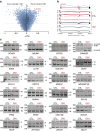
Figure 1
Figure 1. Identification of novel splicing misregulations in DM1 heart samples.
(a) Δ-PSI versus Z-score plot of exon cassettes misregulations predicted by MISO analysis. (b) Exons structure and coverage of RNA-seq reads across SCN5A exons 5–7 show increased inclusion of exon 6A and decreased inclusion of exon 6B in heart samples of three DM1 patients (bottom, blue) versus three control samples (top, red). (c) Validation by RT–PCR of RNA-seq predictions in human heart samples of normal adult individuals (CTL, black) versus adult DM1 patients (DM1, red). Molecular size markers in bps are reported to the left of each RT–PCR gels. bp, base pair.
Figure 2
Figure 2. MBNL-binding motifs are enriched in vicinity of exons misregulated in DM1.
Sequence and binomial test P values of 4-mer RNA motifs enriched downstream, within and upstream of exons misregulated in DM1 heart samples. Sequences enriched in exons excluded in DM are indicated in red, while sequences enriched in exons included in DM are indicated in blue.
Figure 3
Figure 3. Splicing of SCN5A exon 6A is altered in DM heart samples.
(a) Schematic representation of mutually exclusive exons 6A and 6B of SCN5A. SCN5A mRNA includes exon 6A (red) in fetal heart, while SCN5A mRNA expresses exon 6B (blue) in adult heart. (b) Schematic representation of SCN5A topology expressing exon 6A (red). Exons 6A or 6B encodes part of segment 3, connecting loop between S3 and S4 and most part of the voltage-sensitive segment 4 of domain 1 of the sodium channel SCN5A. (c). Representative BstBI-digested RT–PCR analysis of endogenous SCN5A mRNA from human heart samples of normal adult (CTL), adult ALS, non-DM fetuses (20, 24 and 35 weeks), congenital DM1 fetuses (CDM1 of 22, 25 and 28 weeks), adults DM1 and DM2 individuals. Molecular size marker is indicated in bp. (d) Graphical representation of RT–PCR analysis depicting the percentage of SCN5A mRNA including exon 6A in left ventricular heart samples from fetal and adult control, ALS, DCM, DMD, CDM1 and adult DM1 and DM2 individuals. (e) Graphical representation of quantitative real-time RT-qPCR depicting the mRNA expression of SCN5A relative to RPLP0 in control normal adults (n=5) versus adult DM1 (n=5) heart samples. Bars indicate s.e.m. bp, base pairs.
Figure 4
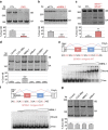
Figure 4. MBNL1 regulates alternative splicing of SCN5A.
(a) Upper panel, RT–PCR analysis of endogenous SCN5A mRNA from differentiated primary muscle cell cultures derived from biopsies of control or DM1 individuals. (lower) Quantification of the percentage of SCN5A mRNA including exon 6B. (b, upper) RT–PCR analysis of endogenous SCN5A mRNA from human differentiated cultures of control primary muscle cells transfected with a scrambled siRNA (siCTL) or a siRNA targeting MBNL1 mRNA (siMBNL1). (lower) Percentage of SCN5A mRNA including exon 6B. (c, upper) RT–PCR analysis of endogenous Scn5a mRNA in heart samples of wild-type and compound Mbnl1−/−, Mbnl2+/− double knockout mice. (lower) Percentage of Scn5a mRNA including exon 6A. (d, upper) RT–PCR analysis of exogenous SCN5A mRNA from differentiated C2C12 muscle cells co-transfected with a SCN5A minigene containing exons 6A and 6B bordered by their introns and with either a plasmid expressing 960 CTG repeats, MBNL1, CUGBP1 or with a siRNA directed against Mbnl1 (siMbnl1) or Celf1 (encoding Cugbp1; siCelf1). # Indicates usage of a cryptic splice site inherent to the minigene. (lower) Percentage of SCN5A mRNA including exon 6B. (e, upper) Schematic representation of SCN5A minigene, including the UGC-rich sequence used for binding assays. (lower) Gel-shift assays were performed using 5–1,000 nM of purified bacterial recombinant GST-MBNL1Δ101 and a uniformly 32P-CTP labelled RNA. (f, upper) Schematic representation of mutant SCN5A minigene, including the mutant sequence, used for binding assays. (lower) Gel-shift assay performed as in e. (g, upper) RT–PCR analysis of exogenous SCN5A mRNA from differentiated C2C12 muscle cells co-transfected with mutant SCN5A minigene and with a plasmid expressing 960 CTG repeats or MBNL1 or with a siRNA directed against Mbnl1 (siMbnl1). (lower) Percentage of SCN5A mRNA including exon 6B. All transfection and gel-shift experiments were repeated three to five times. Molecular size markers are indicated in bp. Bars indicate s.e.m. Student test, ** indicates P<0.01, *** indicates P<0.001. bp, base pairs.
Figure 5
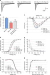
Figure 5. Electrophysiological properties of hNav1.5 and hNav1.5e channels.
(a) Representative Na+ currents generated in Xenopus oocytes by hNav1.5 (encoded by SCN5A containing the adult exon 6B), hNav1.5e (encoded by SCN5A including the fetal exon 6A), and simultaneously expressed Nav1.5 and Nav1.5e channels at a 1:1 ratio. (b) Peak current amplitudes at the test potential of −10 mV in Xenopus oocytes injected with equimolar amount of cRNA encoding hNav1.5, hNav1.5e or 1:1 combination of Nav1.5 and Nav1.5e channels. (c) Current–voltage relationships. (d) Steady-state activation curves. (e) Inactivation time constants τh (ms) at different test pulses. (f) Steady-state inactivation curves. (g) Fractional recovery curves. Data were obtained from 11 different batches of oocytes. To illustrate steady-state activation, steady-state inactivation and recovery from inactivation, we used 3–5 representative measurements. For total number of measurements (n=25–27) and for statistical data evaluation (Vm, s) see the Supplementary Table 2. Bars indicate s.e.m. Student test, *** indicates P<0.001.
Figure 6
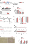
Figure 6. Alteration of Scn5a splicing causes heart conduction defects and arrhythmias.
(a) Schematic representation of mutually exclusive exons 6A and 6B of Scn5a and of antisense sequences driven by optimized U7-snRNAs (U7-ASScn5a) to force fetal exon 6A inclusion in adult wild-type mouse heart. (b, upper) RT–PCR analysis of the alternative splicing of endogenous Scn5a mRNA from heart samples of mice injected with AAV2/9 expressing U7-ASScn5a compared with control injected mice. Molecular size marker is indicated in bp. (lower) Percentage of Scn5a mRNA including exon 6A. (c) Real-time RT-qPCR quantification of the expression of Scn5a, Scn1b and GJja1 (connexin 43) mRNAs in heart samples of mice expressing U7-ASScn5a (n=6) compared with control injected mice (n=6). (d) Representative ECG traces show prolongation of the PR interval in U7-ASScn5a-injected mice compared with control mice. (e) ECG measures of PR interval, QRS and QT intervals in 4-month-old mice injected with AAV2/9 expressing U7-ASScn5a (n=25) compared with age-matched control mice (n=17). (f) Representative ECG traces reveal atrial fibrillation in U7-ASScn5a-injected mice compared with control mice. (g) Variation of the RR interval indicates evidences of heart arrhythmias in U7-ASScn5a-injected mice (n=25) compared with control mice (n=17). (h) Representative image of six analysed heart samples showing mild fibrosis revealed by Red Sirius histology staining in AAV-U7-ASScn5a-injected mice. Scale bar, 100 μm. (i) Real-time RT-qPCR quantification of the expression of Cola1a, Col3a1 and Tgfb mRNAs in heart of control (n=6) or AAV-U7-ASScn5a-injected mice (n=6). Bars indicate s.e.m. Student test, * indicates P<0.5, ** indicates P<0.01, *** indicates P<0.001. bp, base pair.
Figure 7
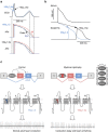
Figure 7. Computer simulation predicts cardiac conduction abnormalities in DM.
(a) Simulations of ECG (upper) and action potential (AP) alterations (middle and lower) caused by the switch from adult SCN5A exon 6B towards fetal exon 6A, using a modified human ventricular ORd model. (b) Simulations of the atrio-ventricular changes caused by inclusion of SCN5A fetal exon 6A instead of adult exon 6B, employing an atrio-ventricular nodal model. APs of atrial and AV node cells were simulated and atrium-His interval was measured as the difference in the latency of APs between atrial and nodal-His cells. (c) Model of splicing alteration of the cardiac sodium channel, SCN5A, in DM. MBNL proteins regulate the switch from SCN5A exon 6A in fetal heart to exon 6B in adult. In DM, titration of MBNL proteins by mutant RNA containing expanded CUG repeats leads to expression of a fetal splicing form of SCN5A, inappropriate to adult heart physiology, ultimately resulting in cardiac-conduction delay and heart arrhythmias, which are two keys features of DM.

References

    1. Brook J. D. et al. Molecular basis of myotonic dystrophy: expansion of a trinucleotide (CTG) repeat at the 3' end of a transcript encoding a protein kinase family member. Cell 68, 799–808 (1992). - PubMed
    1. Fu Y. H. et al. An unstable triplet repeat in a gene related to myotonic muscular dystrophy. Science 255, 1256–1258 (1992). - PubMed
    1. Mahadevan M. et al. Myotonic dystrophy mutation: an unstable CTG repeat in the 3' untranslated region of the gene. Science 255, 1253–1255 (1992). - PubMed
    1. Liquori C. L. et al. Myotonic dystrophy type 2 caused by a CCTG expansion in intron 1 of ZNF9. Science 293, 864–867 (2001). - PubMed
    1. Miller J. W. et al. Recruitment of human muscleblind proteins to (CUG)n expansions associated with myotonic dystrophy. EMBO J. 19, 4439–4448 (2000). - PMC - PubMed

Publication types

MeSH terms