

Early cranial ultrasound findings among infants with neonatal encephalopathy in Uganda: an observational study
- PMID: 27064242
- PMCID: PMC4992358
- DOI: 10.1038/pr.2016.77
Early cranial ultrasound findings among infants with neonatal encephalopathy in Uganda: an observational study
Abstract
Background: In sub-Saharan Africa, the timing and nature of brain injury and their relation to mortality in neonatal encephalopathy (NE) are unknown. We evaluated cranial ultrasound (cUS) scans from term Ugandan infants with and without NE for evidence of brain injury.
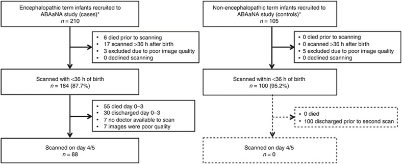
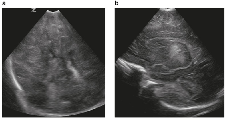
Methods: Infants were recruited from a national referral hospital in Kampala. Cases (184) had NE and controls (100) were systematically selected unaffected term infants. All had cUS scans <36 h reported blind to NE status.
Results: Scans were performed at median age 11.5 (interquartile range (IQR): 5.2-20.2) and 8.4 (IQR: 3.6-13.5) hours, in cases and controls respectively. None had established antepartum injury. Major evolving injury was reported in 21.2% of the cases vs. 1.0% controls (P < 0.001). White matter injury was not significantly associated with bacteremia in encephalopathic infants (odds ratios (OR): 3.06 (95% confidence interval (CI): 0.98-9.60). Major cUS abnormality significantly increased the risk of neonatal death (case fatality 53.9% with brain injury vs. 25.9% without; OR: 3.34 (95% CI: 1.61-6.95)).
Conclusion: In this low-resource setting, there was no evidence of established antepartum insult, but a high proportion of encephalopathic infants had evidence of major recent and evolving brain injury on early cUS imaging, suggesting prolonged or severe acute exposure to hypoxia-ischemia (HI). Early abnormalities were a significant predictor of death.
Figures
References
-
- Lawn JE, Blencowe H, Oza S, et al.; Lancet Every Newborn Study Group. Every Newborn: progress, priorities, and potential beyond survival. Lancet 2014;384:189–205. - PubMed
-
- Rutherford MA, Pennock JM, Dubowitz LM. Cranial ultrasound and magnetic resonance imaging in hypoxic-ischaemic encephalopathy: a comparison with outcome. Dev Med Child Neurol 1994;36:813–25. - PubMed
-
- Cowan F, Rutherford M, Groenendaal F, et al. Origin and timing of brain lesions in term infants with neonatal encephalopathy. Lancet 2003;361:736–42. - PubMed
-
- Martinez-Biarge M, Diez-Sebastian J, Wusthoff CJ, Mercuri E, Cowan FM. Antepartum and intrapartum factors preceding neonatal hypoxic-ischemic encephalopathy. Pediatrics 2013;132:e952–9. - PubMed
Publication types
MeSH terms
Grants and funding
LinkOut - more resources
Full Text Sources
Other Literature Sources
Medical
