

The appearance of newly identified intraocular lesions in Gaucher disease type 3 despite long-term glucocerebrosidase replacement therapy
- PMID: 27064303
- PMCID: PMC4967266
- DOI: 10.3109/03009734.2016.1158756
The appearance of newly identified intraocular lesions in Gaucher disease type 3 despite long-term glucocerebrosidase replacement therapy
Abstract
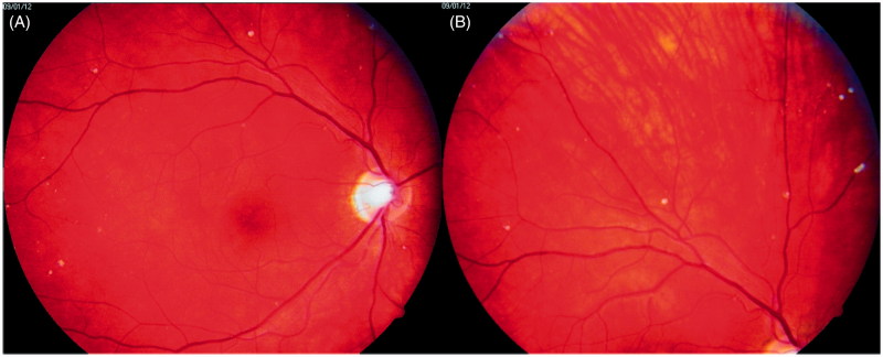
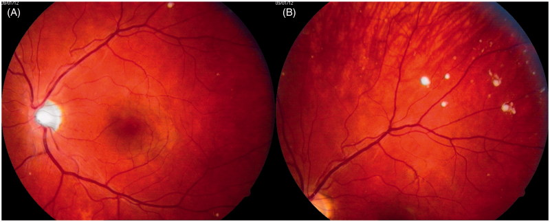
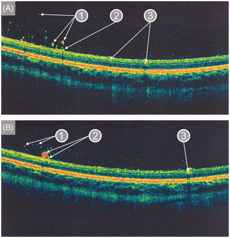
Background Gaucher disease (GD) is an autosomal recessive lipid storage disorder caused by the deficient activity of the lysosomal enzyme glucocerebrosidase. The presence of central nervous system disease is a hallmark of the neuronopathic forms of GD (types 2 and 3). Intraocular lesions (e.g. corneal clouding, retinal lesions, and vitreous opacities) have been infrequently reported in GD type 3 (GD3). Moreover, there are virtually no published data on the occurrence and natural course of intraocular lesions in GD3 patients treated with enzyme replacement therapy (ERT). Case presentation We describe the case of a 26-year-old Polish male with L444P homozygous GD3 (mutation c.1448T > C in the GBA1 gene) who developed fundus lesions despite 10 years of ERT. At the age of 23 years, a spectral domain optical coherence tomography (OCT) examination was performed which disclosed the presence of discrete lesions located preretinally, intraretinally in the nerve fiber layer, and in the vitreous body. A 3-year follow-up OCT examination has not shown any significant progression of the fundus lesions. Conclusions To the best of our knowledge, this is the first published report describing the occurrence of newly identified retinal and preretinal lesions occurring during long-term ERT in GD3. We recommend that a careful ophthalmic assessment, including a dilated fundus examination, should be included as part of annual follow-up in patients with GD3. Further studies are needed to understand the nature and clinical course of these changes and whether or not these intraocular findings have any predictive value in the context of neurologic and skeletal progression in GD3.
Keywords: Enzyme replacement therapy; Gaucher disease type 3; intraocular lesions; neuronopathic; optical coherence tomography; retina.
Figures
References
-
- Zimran A. How I treat Gaucher disease. Blood 2011;118:1463–71. - PubMed
-
- Hruska KS, LaMarca ME, Scott CR, Sidransky E.. Gaucher disease: mutation and polymorphism spectrum in the glucocerebrosidase gene (GBA). Hum Mutat 2008;29:567–83. - PubMed
-
- Machaczka M, Markuszewska-Kuczyńska A, Regenthal S, Jurczyszyn A, Gałązka K, Wahlin BE, et al. Clinical utility of different bone marrow examination methods in the diagnosis of adults with sporadic Gaucher disease type 1. Pol Arch Med Wewn 2014;124:587–92. - PubMed
-
- Markuszewska-Kuczyńska A, Klimkowska M, Regenthal S, Bulanda A, Kämpe Björkvall C, Machaczka M.. Atypical cytomorphology of Gaucher cells is frequently seen in bone marrow smears from untreated patients with Gaucher disease type 1. Folia Histochem Cytobiol 2015;53:62–9. - PubMed
-
- Tylki-Szymańska A, Vellodi A, El-Beshlawy A, Cole JA, Kolodny E.. Neuronopathic Gaucher disease: demographic and clinical features of 131 patients enrolled in the International Collaborative Gaucher Group Neurological Outcomes Subregistry. J Inherit Metab Dis 2010;33:339–46. - PubMed
Publication types
MeSH terms
Substances
LinkOut - more resources
Full Text Sources
Other Literature Sources
Medical