Review
. 2016 Mar 15;6(2):561-601.
doi: 10.1002/cphy.c140071.

PTH and Vitamin D

Affiliations
Review

PTH and Vitamin D

Syed Jalal Khundmiri et al. Compr Physiol. .

Abstract

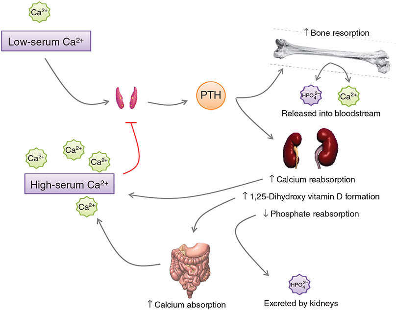
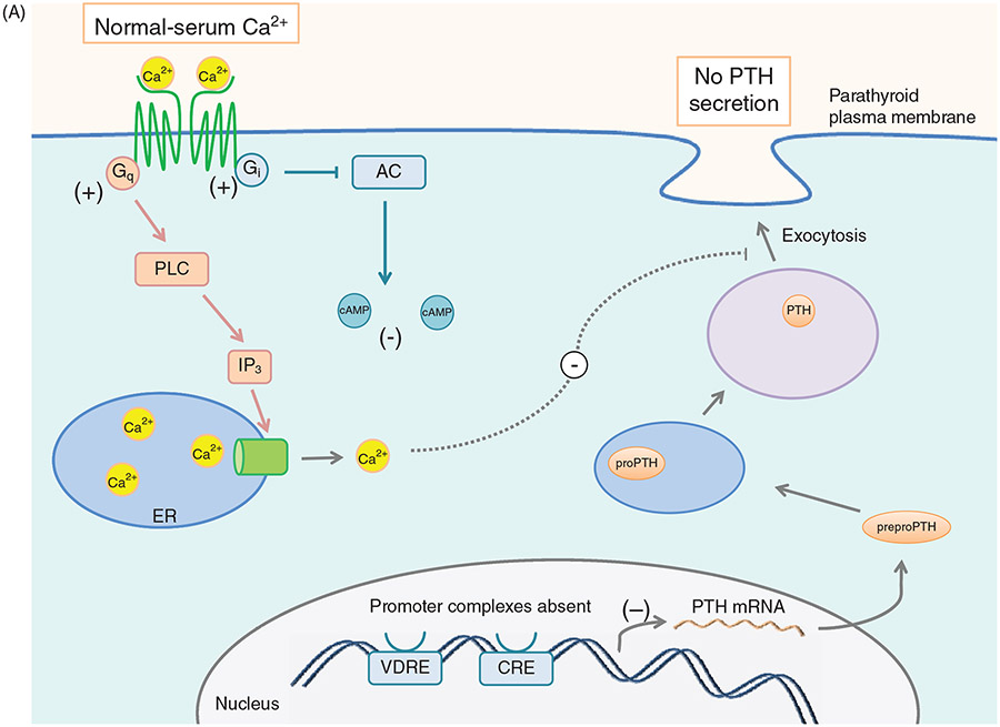
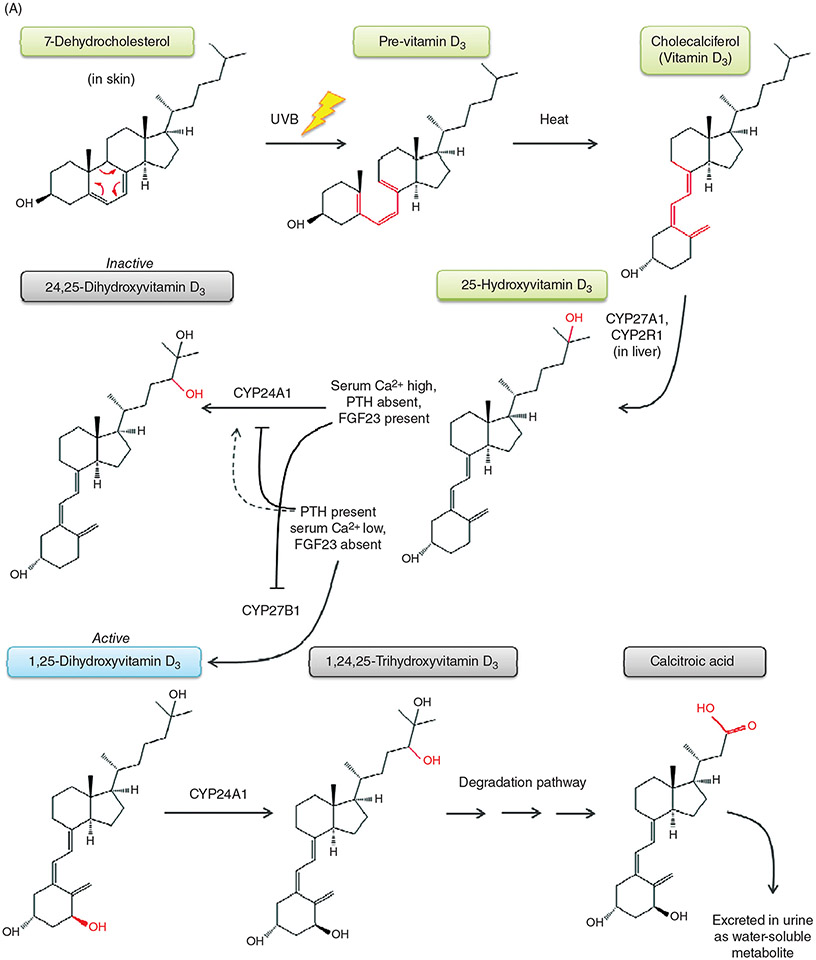
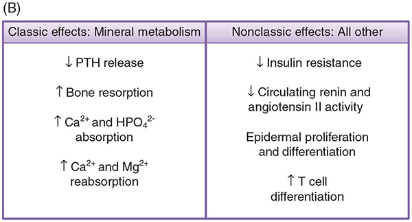
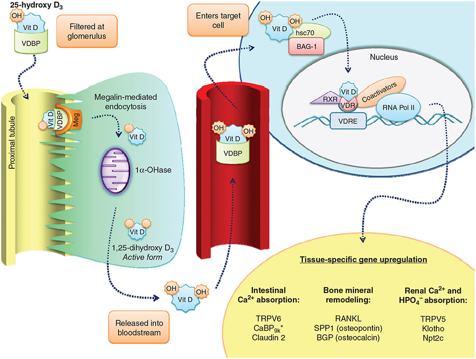
PTH and Vitamin D are two major regulators of mineral metabolism. They play critical roles in the maintenance of calcium and phosphate homeostasis as well as the development and maintenance of bone health. PTH and Vitamin D form a tightly controlled feedback cycle, PTH being a major stimulator of vitamin D synthesis in the kidney while vitamin D exerts negative feedback on PTH secretion. The major function of PTH and major physiologic regulator is circulating ionized calcium. The effects of PTH on gut, kidney, and bone serve to maintain serum calcium within a tight range. PTH has a reciprocal effect on phosphate metabolism. In contrast, vitamin D has a stimulatory effect on both calcium and phosphate homeostasis, playing a key role in providing adequate mineral for normal bone formation. Both hormones act in concert with the more recently discovered FGF23 and klotho, hormones involved predominantly in phosphate metabolism, which also participate in this closely knit feedback circuit. Of great interest are recent studies demonstrating effects of both PTH and vitamin D on the cardiovascular system. Hyperparathyroidism and vitamin D deficiency have been implicated in a variety of cardiovascular disorders including hypertension, atherosclerosis, vascular calcification, and kidney failure. Both hormones have direct effects on the endothelium, heart, and other vascular structures. How these effects of PTH and vitamin D interface with the regulation of bone formation are the subject of intense investigation.


Figures

Figure 1
Figure 1
Regulation of serum Ca2+ by PTH. Low serum Ca2+ stimulates the release of PTH from the parathyroid gland. PTH acts on the bone to increase bone resorption, releasing Ca2+ and HPO42− into the bloodstream. At the kidney, PTH increases Ca2+ reabsorption and decreases HPO42− reabsorption, maintaining the elevated serum Ca2+ from the resorption of bone. Vitamin D becomes activated in the kidney by 1α-hydroxylase, leading to increased Ca2+ absorption from the gut. The restored serum Ca2+ provides a negative feedback signal to the parathyroid glands, discontinuing the release of PTH.
Figure 2
Figure 2
Primary sequence of human parathyroid hormone (sequence from Ensembl.org). The pre- and prosequences are cleaved prior to secretion into the bloodstream. Residues 1 and 2 (diagonally striped) are required for PTH receptor activation. Residues 28-34 (checkered pattern) interact with the extracellular amino domain of the PTH receptor. The C-fragment may be cleaved either prior to or after release from the parathyroid gland.
Figure 3
Figure 3
(A) Effect of serum [Ca2+] on PTH synthesis and secretion. Ca2+ binds to the CaSR and activates Gq and Gi. Gq activation leads to increased [Ca2+]i via PLC signaling and IP3 formation. Increased [Ca2+]i inhibits exocytosis of PTH secretory vesicles. Gi activation inhibits adenylyl cyclase, leading to less cAMP, and therefore less transcription of the PTH gene as well as decreased stimulus for secretory vesicle exocytosis. (B) Effect of low serum [Ca2+] on PTH synthesis and secretion. In the absence of serum Ca2+, CaSR signaling terminates, allowing [cAMP]i to increase, which drives transcription of PTH and secretory vesicle exocytosis, leading to increased serum PTH levels. (C) Effect of Vitamin D on PTH secretion. Vitamin D enters the cell through diffusion across the plasma membrane. Once inside, Vitamin D is bound by the vitamin D receptor (VDR). In the nucleus, the Vit D-VDR complex binds to RXR and the vitamin D response element (VDRE) within the PTH promoter. This promoter complex inhibits transcription of the PTH gene, leading to less preproPTH production. The Vit D-VDR complex also binds to the VDRE within the promoter of p21, activating the transcription of the p21 gene. Increased p21 expression inhibits parathyroid cell proliferation.
Figure 3
Figure 3
(A) Effect of serum [Ca2+] on PTH synthesis and secretion. Ca2+ binds to the CaSR and activates Gq and Gi. Gq activation leads to increased [Ca2+]i via PLC signaling and IP3 formation. Increased [Ca2+]i inhibits exocytosis of PTH secretory vesicles. Gi activation inhibits adenylyl cyclase, leading to less cAMP, and therefore less transcription of the PTH gene as well as decreased stimulus for secretory vesicle exocytosis. (B) Effect of low serum [Ca2+] on PTH synthesis and secretion. In the absence of serum Ca2+, CaSR signaling terminates, allowing [cAMP]i to increase, which drives transcription of PTH and secretory vesicle exocytosis, leading to increased serum PTH levels. (C) Effect of Vitamin D on PTH secretion. Vitamin D enters the cell through diffusion across the plasma membrane. Once inside, Vitamin D is bound by the vitamin D receptor (VDR). In the nucleus, the Vit D-VDR complex binds to RXR and the vitamin D response element (VDRE) within the PTH promoter. This promoter complex inhibits transcription of the PTH gene, leading to less preproPTH production. The Vit D-VDR complex also binds to the VDRE within the promoter of p21, activating the transcription of the p21 gene. Increased p21 expression inhibits parathyroid cell proliferation.
Figure 4
Figure 4
(A) PTH receptor topology and ligand binding. (A)The PTH1R consists of an extracellular amino terminus, a J domain consisting of transmembrane domains as well as intra- and extracellular loops, and an intracellular carboxy terminus. The extracellular amino terminus is 150 residues long, with 4 N-glycosylation sites and 4 disulfide bridges. Less is known regarding PTH2R topology as compared to PTH1R topology. Importantly, residue variations in two of the extracellular loops (highlighted in red) decrease the affinity of the receptor for PTHrP while maintaining specificity for PTH. (B) PTH receptor topology and ligand binding. (B) PTH(1-34) (rainbow structure) interacts with both the extracellular amino terminus of the PTH1R as well as the J domain. Docking of PTH to the PTH1R is thought to occur through initial binding of the C-terminus of PTH (residues 15-34) to the N-terminus (blue regions) of the PTH1R. This interaction is closely followed by the binding of the N-terminus of PTH (residues 1-14) to the J domain (red regions) of the PTH1R, initiating G protein recruitment and intracellular signaling cascade activation. (C) PTH-receptor binding and intracellular signaling. (1) Two-site model of PTH-receptor docking. (2) The N-domain of the PTH receptor (PTHR1) binds the C-domain of PTH. (3) The J-domain binds the amino-terminal region of PTH. (4) Binding of the ligand to the receptor increases the association with the J-domain, while also increasing the affinity of the intracellular beta-gamma binding region of the C-terminal region of the PTH receptor for G proteins, resulting in their subsequent activation and initiation of downstream signaling cascades. Gαs activates adenylate cyclase (AC), which increases intracellular [cAMP], resulting in activation of Epac and PKA. Gq activates phospholipase C (PLC), forming diacylglycerol (DAG) and inositol triphosphate (IP3). DAG directly activates PKC, whereas IP3 indirectly activates PKC by releasing Ca2+ from the ER.
Figure 4
Figure 4
(A) PTH receptor topology and ligand binding. (A)The PTH1R consists of an extracellular amino terminus, a J domain consisting of transmembrane domains as well as intra- and extracellular loops, and an intracellular carboxy terminus. The extracellular amino terminus is 150 residues long, with 4 N-glycosylation sites and 4 disulfide bridges. Less is known regarding PTH2R topology as compared to PTH1R topology. Importantly, residue variations in two of the extracellular loops (highlighted in red) decrease the affinity of the receptor for PTHrP while maintaining specificity for PTH. (B) PTH receptor topology and ligand binding. (B) PTH(1-34) (rainbow structure) interacts with both the extracellular amino terminus of the PTH1R as well as the J domain. Docking of PTH to the PTH1R is thought to occur through initial binding of the C-terminus of PTH (residues 15-34) to the N-terminus (blue regions) of the PTH1R. This interaction is closely followed by the binding of the N-terminus of PTH (residues 1-14) to the J domain (red regions) of the PTH1R, initiating G protein recruitment and intracellular signaling cascade activation. (C) PTH-receptor binding and intracellular signaling. (1) Two-site model of PTH-receptor docking. (2) The N-domain of the PTH receptor (PTHR1) binds the C-domain of PTH. (3) The J-domain binds the amino-terminal region of PTH. (4) Binding of the ligand to the receptor increases the association with the J-domain, while also increasing the affinity of the intracellular beta-gamma binding region of the C-terminal region of the PTH receptor for G proteins, resulting in their subsequent activation and initiation of downstream signaling cascades. Gαs activates adenylate cyclase (AC), which increases intracellular [cAMP], resulting in activation of Epac and PKA. Gq activates phospholipase C (PLC), forming diacylglycerol (DAG) and inositol triphosphate (IP3). DAG directly activates PKC, whereas IP3 indirectly activates PKC by releasing Ca2+ from the ER.
Figure 5
Figure 5
(A) Metabolic pathway of vitamin D. (A) The production of 1,25-dihydroxyvitamin D begins in the skin with the precursor 7-dehydrocholesterol. UVB light catalyzes the conversion of 7-DHC to pre-Vitamin D (structural changes denoted in red), which is further converted to Vitamin D by heat through the process of thermal isomerization. Vitamin D enters the bloodstream, and in the liver undergoes hydroxylation by either CYP27A1 or CYP2R1 to form 25-hydroxyvitamin D. Depending on serum Ca2+ levels and hormones present, in the kidney, 25-hydroxyvitamin D will either be converted to its active form, 1,25-dihydroxyvitamin D, by the enzyme CYP27B1, or be converted to its inactive form, 24,25-dihydroxyvitamin D, by the enzyme CYP24A1. FGF23 upregulates CYP24A1 expression and downregulates CYP27B1. PTH stimulates CYP27B1 expression, and depending on the circumstances, either downregulates CYP24A1 or slightly upregulates its expression. 1,25-dihydroxyvitamin D3 is degraded first by hydroxylation at the 24th position to form 1,24,25-trihydroxyvitamin D, then through a series of steps form the end product calcitroic acid, which is water-soluble and is excreted in the urine. (B) 1,25-Dihydroxyvitamin D3 promotes both classic effects on mineral metabolism as well as nonclassic effects on immune function, cardiovascular protection, and others as listed. (B) Metabolic pathway of vitamin D. (A) The production of 1,25-dihydroxyvitamin D begins in the skin with the precursor 7-dehydrocholesterol. UVB light catalyzes the conversion of 7-DHC to previtamin D, which is further converted to vitamin D by heat. Vitamin D enters the bloodstream, and in the liver undergoes hydroxylation by either CYP27A1 or CYP2R1 to form 25-hydroxyvitamin D. Depending on serum Ca2+ levels and hormones present, in the kidney, 25-hydroxyvitamin D will either be converted to its active form, 1,25-dihydroxyvitamin D, by the enzyme CYP27B1, or be converted to its inactive form, 24,25-dihydroxyvitamin D, by the enzyme CYP24A1. 1,25-dihydroxy-D3 is degraded through a series of steps to the water-soluble product calcitroic acid, which is excreted in the urine. (B) 1,25-Dihydroxy-D3 promotes both classic effects on mineral metabolism as well as nonclassic effects on immune function, cardiovascular protection, and others.
Figure 5
Figure 5
(A) Metabolic pathway of vitamin D. (A) The production of 1,25-dihydroxyvitamin D begins in the skin with the precursor 7-dehydrocholesterol. UVB light catalyzes the conversion of 7-DHC to pre-Vitamin D (structural changes denoted in red), which is further converted to Vitamin D by heat through the process of thermal isomerization. Vitamin D enters the bloodstream, and in the liver undergoes hydroxylation by either CYP27A1 or CYP2R1 to form 25-hydroxyvitamin D. Depending on serum Ca2+ levels and hormones present, in the kidney, 25-hydroxyvitamin D will either be converted to its active form, 1,25-dihydroxyvitamin D, by the enzyme CYP27B1, or be converted to its inactive form, 24,25-dihydroxyvitamin D, by the enzyme CYP24A1. FGF23 upregulates CYP24A1 expression and downregulates CYP27B1. PTH stimulates CYP27B1 expression, and depending on the circumstances, either downregulates CYP24A1 or slightly upregulates its expression. 1,25-dihydroxyvitamin D3 is degraded first by hydroxylation at the 24th position to form 1,24,25-trihydroxyvitamin D, then through a series of steps form the end product calcitroic acid, which is water-soluble and is excreted in the urine. (B) 1,25-Dihydroxyvitamin D3 promotes both classic effects on mineral metabolism as well as nonclassic effects on immune function, cardiovascular protection, and others as listed. (B) Metabolic pathway of vitamin D. (A) The production of 1,25-dihydroxyvitamin D begins in the skin with the precursor 7-dehydrocholesterol. UVB light catalyzes the conversion of 7-DHC to previtamin D, which is further converted to vitamin D by heat. Vitamin D enters the bloodstream, and in the liver undergoes hydroxylation by either CYP27A1 or CYP2R1 to form 25-hydroxyvitamin D. Depending on serum Ca2+ levels and hormones present, in the kidney, 25-hydroxyvitamin D will either be converted to its active form, 1,25-dihydroxyvitamin D, by the enzyme CYP27B1, or be converted to its inactive form, 24,25-dihydroxyvitamin D, by the enzyme CYP24A1. 1,25-dihydroxy-D3 is degraded through a series of steps to the water-soluble product calcitroic acid, which is excreted in the urine. (B) 1,25-Dihydroxy-D3 promotes both classic effects on mineral metabolism as well as nonclassic effects on immune function, cardiovascular protection, and others.
Figure 6
Figure 6
Vitamin D effector mechanisms. 25-Hydroxy-vitamin D bound to DBP is filtered, binds to the megalin complex and is endocytosed into the renal proximal tubule cell where it undergoes 1 α hydroxylation. Activated vitamin D bound to cofactors and chaperones (BAG-1 and hsc70) enter the nucleus and interacts with the VDR in concert with the RXR. This complex interacts with specific genomic regions including the VDRE to influence gene transcription.

References

    1. Abe Y, Nagasaki K, Watanabe T, Abe T, Fukami M. Association between compound heterozygous mutations of SLC34A3 and hypercalciuria. Horm Res Paediatr 82: 65–71, 2014. - PubMed
    1. Adamczak DM, Nowak JK, Frydrychowicz M, Kaczmarek M, Sikora J. The role of Toll-like receptors and vitamin D in diabetes mellitus type 1–a review. Scand J Immunol 80: 75–84, 2014. - PubMed
    1. Adams AE, Bisello A, Chorev M, Rosenblatt M, Suva LJ. Arginine 186 in the extracellular N-terminal region of the human parathyroid hormone 1 receptor is essential for contact with position 13 of the hormone. Mol Endocrinol (Baltimore, Md) 12: 1673–1683, 1998. - PubMed
    1. Adams JS, Chen H, Chun R, Gacad MA, Encinas C, Ren S, Nguyen L, Wu S, Hewison M, Barsony J. Response element binding proteins and intracellular vitamin D binding proteins: NMovel regulators of vitamin D trafficking, action and metabolism. J Steroid Biochem Mol Biol 89-90: 461–465, 2004. - PubMed
    1. Adams JS, Clemens TL, Parrish JA, Holick MF. Vitamin-D synthesis and metabolism after ultraviolet irradiation of normal and vitamin-D-deficient subjects. N Engl J Med 306: 722–725, 1982. - PubMed

Publication types

MeSH terms