Review
. 2016 Mar 15;6(2):897-943.
doi: 10.1002/cphy.c140049.

Lung Circulation

Affiliations
Review

Lung Circulation

Karthik Suresh et al. Compr Physiol. .

Abstract

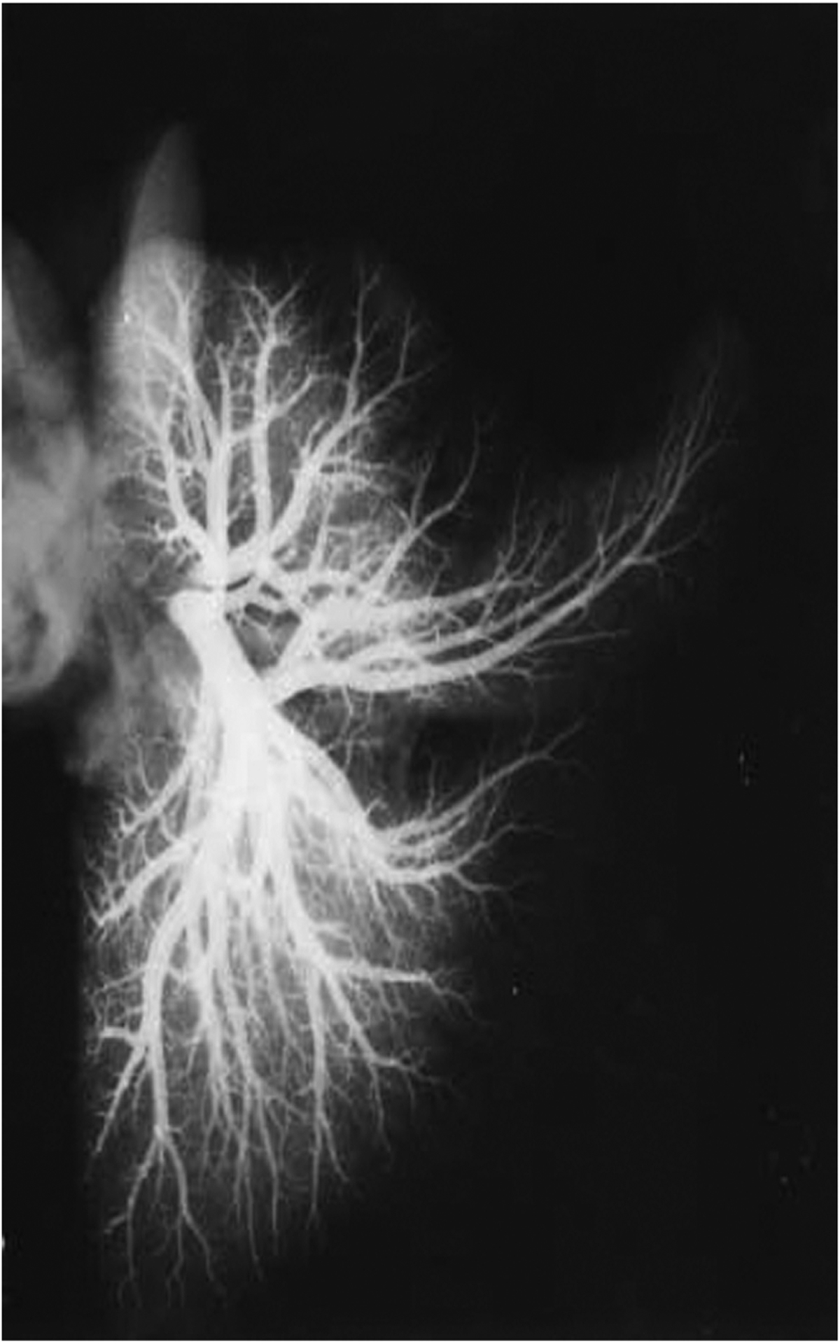
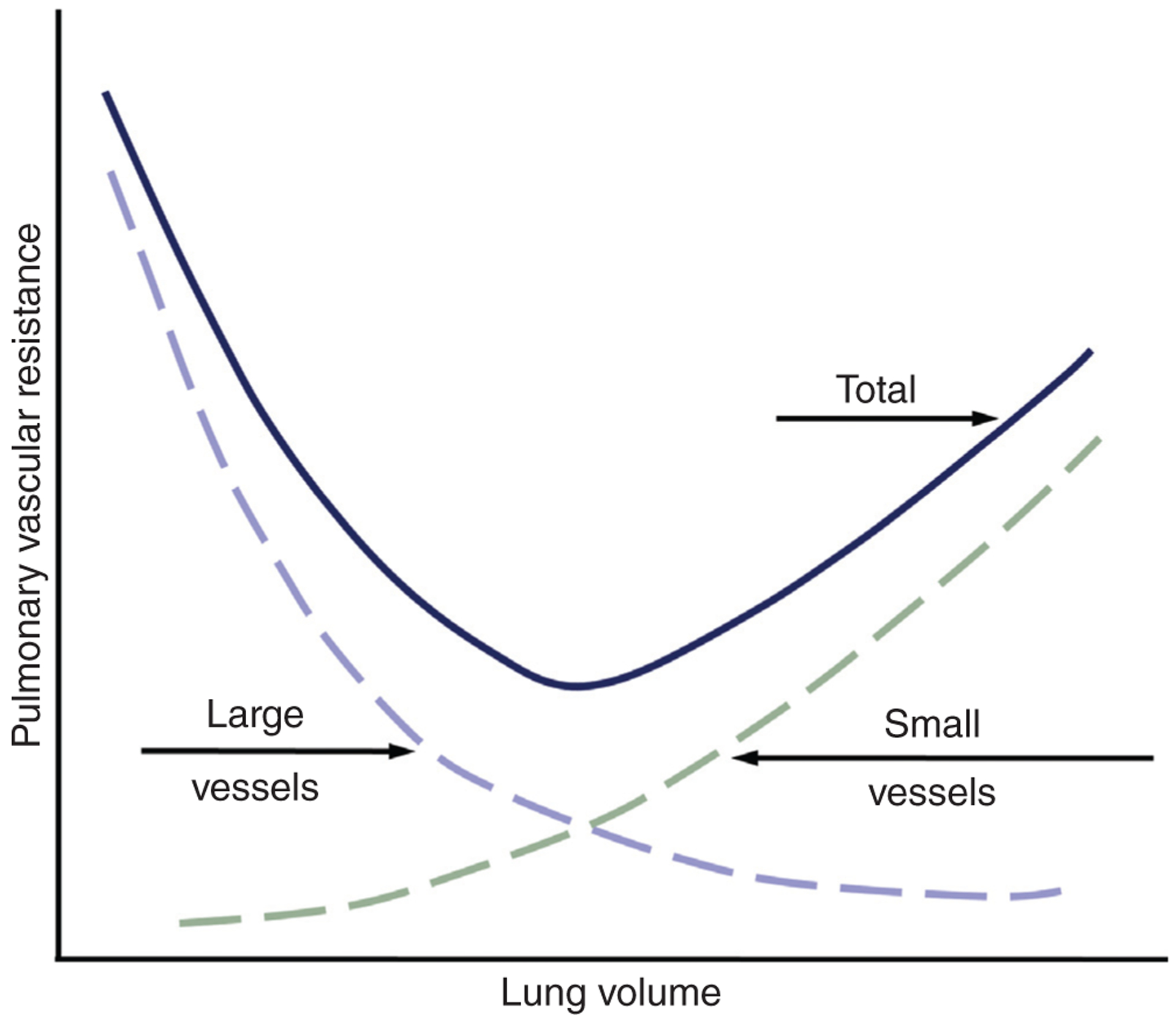
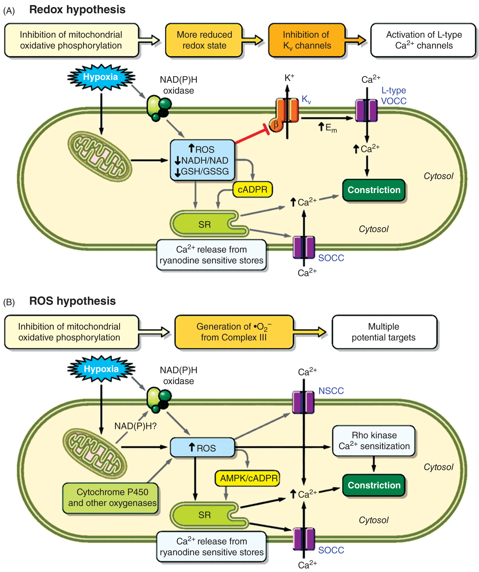
The circulation of the lung is unique both in volume and function. For example, it is the only organ with two circulations: the pulmonary circulation, the main function of which is gas exchange, and the bronchial circulation, a systemic vascular supply that provides oxygenated blood to the walls of the conducting airways, pulmonary arteries and veins. The pulmonary circulation accommodates the entire cardiac output, maintaining high blood flow at low intravascular arterial pressure. As compared with the systemic circulation, pulmonary arteries have thinner walls with much less vascular smooth muscle and a relative lack of basal tone. Factors controlling pulmonary blood flow include vascular structure, gravity, mechanical effects of breathing, and the influence of neural and humoral factors. Pulmonary vascular tone is also altered by hypoxia, which causes pulmonary vasoconstriction. If the hypoxic stimulus persists for a prolonged period, contraction is accompanied by remodeling of the vasculature, resulting in pulmonary hypertension. In addition, genetic and environmental factors can also confer susceptibility to development of pulmonary hypertension. Under normal conditions, the endothelium forms a tight barrier, actively regulating interstitial fluid homeostasis. Infection and inflammation compromise normal barrier homeostasis, resulting in increased permeability and edema formation. This article focuses on reviewing the basics of the lung circulation (pulmonary and bronchial), normal development and transition at birth and vasoregulation. Mechanisms contributing to pathological conditions in the pulmonary circulation, in particular when barrier function is disrupted and during development of pulmonary hypertension, will also be discussed.


Figures

Figure 1
Figure 1
Timeline of vascular development. Adapted, with permission, from Hislop and Pierce (265).
Figure 2
Figure 2
Arteriogram of a human lung obtained postmortem from a fetus (39 weeks gestation) showing distal vascularization. The arteries were injected with barium sulphate (mag × 0.75). Reprinted, with permission, from (265).
Figure 3
Figure 3
Graph illustrating the relationship between PVR and volume. Adapted, with permission, from (581).
Figure 4
Figure 4
Original three-zone model proposed by West et al., (688) to illustrate the regional heterogeneity in blood flow. Within each of the three zones, the behavior of the blood vessels is different, based on the relative magnitudes of pulmonary arterial, alveolar and venous pressures (Pa, PA, and Pv, respectively). In zone 1, PA is greater than Pa, occluding collapsible vessels and preventing flow. In zone 2, Pa is greater than PA, which exceeds Pv, such that blood flow is dictated by the Pa-PA pressure gradient. In zone 3, both Pa and Pv exceed PA, and vessels are held open, allowing blood flow based on the Pa-Pv pressure gradient. Reproduced, with permission, from West et al. (688).
Figure 5
Figure 5
Effect of PEEP and no, or zero, end expiratory pressure on VR and CO. (A) Effect of PEEP on CO and VR if PEEP has no effects on venous return flow limitation. Points A and B represent effects of PEEP without changes in mean systemic pressure (Pms). Point C represents effect of PEEP with Pms compensation. (B) Effect of PEEP on CO and venous return if increased VR flow limitation (FL2) occurs with higher PEEP. Reprinted, with permission, from Luecke and Pelosi (381).
Figure 6
Figure 6
Proposed mechanisms by which hypoxia caused pulmonary vasoconstriction. Reproduced, with permission, from Sylvester et al. (606).
Figure 7
Figure 7
The lung-on-a-chip microdevice. (A) Compartmentalized channels form an alveolar-capillary barrier on a thin, porous, flexible membrane. Physiological breathing movements are mimicked by applying a vacuum to the side chambers, causing mechanical stretching of the membrane that forms the alveolar-capillary barrier. (B) Cartoon illustrating the mechanical effects of inspiration in the living lung, where contraction of the diaphragm reduced pleural pressure (Pip), resulting in distension of alveoli and physical stretching of the alveolar-capillary interface. (C) The three-layer system forming parallel microchannels with a porous membrane. Scale bar, 200 μm. (D) After the three layers are bonded together, the membrane layers in the side channels are removed, producing two side chambers to which vacuum is applied to cause mechanical stretching. Scale bar, 200 μm. (E) Actual images of a lung-on-a-chip device. Reproduced, with permission, from (279).
Figure 8
Figure 8
Illustration of putative mechanisms involved in the pathogenesis of pulmonary hypertension. Abbreviations: 5-HT, 5-hydroxytryptamin; K- and Ca-channels, potassium and calcium channels; AEC, alveolar epithelial cells; BMP, bone morphogenetic protein; cGMP, cyclic guanosine monophosphate; ECM, extracellular matrix; EGF, epidermal growth factor; EPC, endothelial progenitor cells; HIF, hypoxia inducible factor; MMPs, matrix metalloproteinases; NADPH, nicotinamide adenine dinucleotide phosphate; NO, nitric oxide; PDE, phosphodiesterase; PDGF, platelet-derived growth factor; PGI2, prostaglandin I2; Rho-Ki, Rho kinases; ROS, reactive oxygen species; sGC, soluble guanylate cyclase; TGF, transforming growth factor-β; TK, tyrosine kinase; TKi, tyrosine kinase inhibitor; TRPC, transient receptor potential cation channels; VEGF, vascular endothelial growth factor. Reproduced, with permission, from Schermuly et al. (549).
Figure 9
Figure 9
Drawing by Fredrick Ruysch detailing the bronchial circulation. Reproduced, with permission, from Mitzner (424).

Similar articles

Cited by

References

    1. Aaronson PI, Robertson TP, Knock GA, Becker S, Lewis TH, Snetkov V, Ward JP. Hypoxic pulmonary vasoconstriction: mechanisms and controversies. J Physiol 570: 53–58, 2006. - PMC - PubMed
    1. Abe K, Shimokawa H, Morikawa K, Uwatoku T, Oi K, Matsumoto Y, Hattori T, Nakashima Y, Kaibuchi K, Sueishi K, Takeshit A. Long-term treatment with a Rho-kinase inhibitor improves monocrotaline-induced fatal pulmonary hypertension in rats. Circ Res 94: 385–393, 2004. - PubMed
    1. Abe K, Toba M, Alzoubi A, Ito M, Fagan KA, Cool CD, Voelkel NF, McMurtry IF, Oka M. Formation of plexiform lesions in experimental severe pulmonary arterial hypertension. Circulation 121: 2747–2754, 2010. - PubMed
    1. Abe T, Arakawa Y, Rajasekaran AK, Yu TH, Wada O. Interaction of atrial natriuretic peptide with its receptors in bovine lung membranes. J Biol Chem 270: 7672–7678, 1995. - PubMed
    1. Abman SH, Accurso FJ. Acute effects of partial compression of ductus arteriosus on fetal pulmonary circulation. Am J Physiol 257: H626–H634, 1989. - PubMed

Publication types