. 2016 Oct 27;35(43):5663-5673.
doi: 10.1038/onc.2016.103. Epub 2016 Apr 11.

Elimination of chronic lymphocytic leukemia cells in stromal microenvironment by targeting CPT with an antiangina drug perhexiline

Affiliations

Elimination of chronic lymphocytic leukemia cells in stromal microenvironment by targeting CPT with an antiangina drug perhexiline

P-P Liu et al. Oncogene. .

Abstract

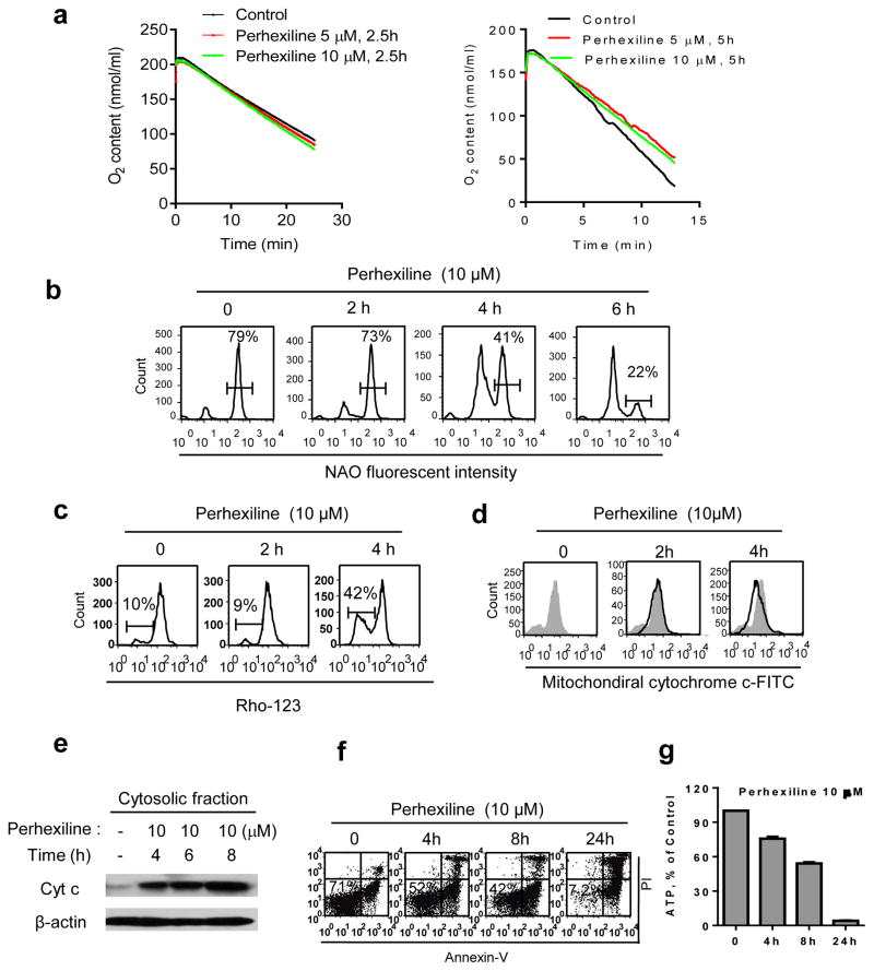
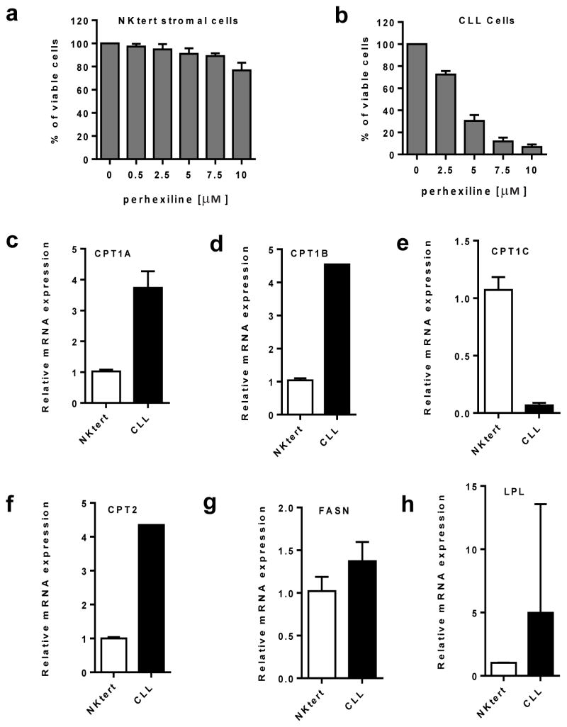
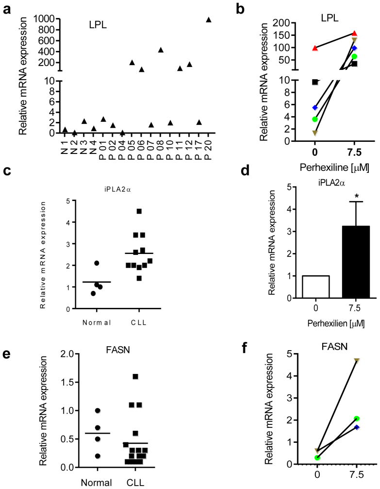
Chronic lymphocytic leukemia (CLL) is the most common adult leukemia in the western countries and is currently incurable due, in part, to difficulty in eliminating the leukemia cells protected by stromal microenvironment. Based on previous observations that CLL cells exhibit mitochondrial dysfunction and altered lipid metabolism and that carnitine palmitoyltransferases (CPT) have a major role in transporting fatty acid into mitochondria to support cancer cell metabolism, we tested several clinically relevant inhibitors of lipid metabolism for their ability to eliminate primary CLL cells. We discovered that perhexiline, an antiangina agent that inhibits CPT, was highly effective in killing CLL cells in stromal microenvironment at clinically achievable concentrations. These effective concentrations caused low toxicity to normal lymphocytes and normal stromal cells. Mechanistic study revealed that CLL cells expressed high levels of CPT1 and CPT2. Suppression of fatty acid transport into mitochondria by inhibiting CPT using perhexiline resulted in a depletion of cardiolipin, a key component of mitochondrial membranes, and compromised mitochondrial integrity, leading to rapid depolarization and massive CLL cell death. The therapeutic activity of perhexiline was further demonstrated in vivo using a CLL transgenic mouse model. Perhexiline significantly prolonged the overall animal survival by only four drug injections. Our study suggests that targeting CPT using an antiangina drug is able to effectively eliminate leukemia cells in vivo, and is a novel therapeutic strategy for potential clinical treatment of CLL.


Conflict of interest statement

The authors declare no conflict of interest

Figures

Figure 1
Figure 1
Identification of perhexiline as a potent drug that effectively killed CLL cells in the presence of bone marrow stromal cells. (a) Schematic illustration of major lipid metabolic pathways and the target enzymes (green) of the three drugs (yellow) examined in this study. TG, triacylglycerol; LPL, lipoprotein lipase; FA, fatty acid; LPA, lysophosphatidic acid; PA, phosphatidic acid; PGP, phosphatidylglycerophosphate; CL, cardiolipin; LCFA-CoAs, long-chain fatty acyl Coenzyme A; FASN, fatty acid synthase; CPT, carnitine palmitoyl transferase; TCA cycle, tricarboxylic-acid cycle. (b) Primary CLL cells were incubated with various concentrations of cerulenin (FASN inhibitor) for 48 h in the presence and absence of StromaNKtert cells as indicated, and cell viability was measured by annexin-V/PI staining and flow cytometry analysis. The results from a representative CLL sample are shown on the left panels. The number (%) within each flow cytometry panel indicates % of viable cells (annexin-V/PI double-negative). Quantitative results of 3 CLL patient samples are shown on the right panel. Each bar shows mean ± SD of three separate experiments with 3 patient samples. (c) Primary CLL cells were incubated with various concentrations of ranolazine (β-oxidation inhibitor) for 48 h in the presence and absence of StromaNKtert cells as indicated, and cell viability was measured as described in b, n=3 patient samples. (d) Primary CLL cells were incubated with various concentrations of pehexiline for 48 h in the presence and absence of StromaNKtert cells as indicated, and cell viability was measured as in described in b. n=19 patient samples. In the co-culture experiments, the ratio of stromal cells:CLL cells was 1:25. In the control experiments in b, c, and d, cells were incubated with solvent (0.1% DMSO) for 48 h, which was not toxic to CLL cells as shown in Supplementary Figure S2.
Figure 2
Figure 2
Effect of perhexiline on cardiolipin and mitochondrial membrane integrity in CLL cells. (a) Primary CLL cells were treated with or without 5–10 μM perhexiline as indicated. CLL cells were suspended in fresh medium (108 cells/ml) and oxygen consumption was measured using an Oxythem system as described under Method. Data were representative of experiments using two separate patient samples. (b) Rapid depletion of cardiolipin induced by perhexiline. CLL cells were incubated with 10 μM perhexiline as indicated, and cardiolipin contents were measured using NAO staining and flow cytometry analysis. The number (%) in each panel indicates the % of cells with normal cardiolipin content. Data were representative of experiments using 3 separate patient samples. (c) Induction of mitochondrial transmembrane potential loss by 10 μM perhexiline in primary CLL cells, detected by flow cytometry analysis using Rho-123 staining. The % cells that lost transmembrane potential are indicated by the number (%) within each panel. Data were representative of experiments using 3 separate patient samples. (d) Loss of mitochondrial cytochrome c induced by perhexiline (10 μM) in CLL cells. Mitochondrial cytochrome c was measured by flow cytometry analysis as described under Method. The control sample is shown in gray (shaded) and the perhexiline-treated samples are shown in black curves. Data were representative of experiments using 3 patient samples. (e) Western blot analysis of cytosolic cytochrome c released from mitochondria before and after perhexiline treatment. CLL cells were incubated with 10 μM perhexiline for the indicated times, and cytosolic proteins were isolated for western blot analysis of cytochrome c release from mitochondria. (f) Time-dependent cell death induced by 10 μM perhexiline. Cell viability was analyzed by flow cytometry after cells were double stained with annexin-V/PI. G, primary CLL cells were incubated with 10 μM perhexiline for 4–24 h as indicated; 5x10⁵ cells from each sample were collected for ATP analysis as described under Method, n=3 patient samples.
Figure 3
Figure 3
Selective killing of CLL cells by perhexiline in comparison with normal lymphocytes. (a) Primary CLL cells or normal lymphocytes were treated with or without 5 μM perhexiline for 48 h, and cell viability was analyzed by annexin-V/PI staining. The number (%) within each panel indicates % of viable cells (Annexin-V/PI double-negative). The quantitative results of 19 CLL patient samples and normal lymphocytes from 5 healthy donors are shown on the right panel. Each bar shows mean ± SD; **, P<0.01. (b) CLL cells or normal lymphocytes were treated with or without 5 μM perhexiline in the presence of bone marrow stromal cells (StromaNKtert) for 48 h. Cell viability was analyzed annexin-V/PI staining. The quantitative results of 19 CLL patient samples and 5 normal lymphocyte samples are shown on the right panel. Each bar shows mean ± SD; **, P <0.01. (c) Normal lymphocytes from healthy donors (n=3) were incubated with 10 μM perhexiline for 2–6 h as indicated. Cardiolipin was measured by flow cytometry using NAO staining. The number within each panel indicates % of cells with normal NAO fluorescent signal. (d) Normal lymphocytes (n=3) were incubated with 10 μM perhexiline for 2–4 h as indicated; mitochondrial transmembrane potential was then measured by flow cytometry after cells were stained with Rho-123. The number within each panel indicates % of cells that lost transmembrane potential. (e) Comparison of sensitivity of CLL cells and normal lymphocytes to perhexiline. CLL cells isolated from 11 CLL patients or normal lymphocytes from 5 healthy donors were incubated with the indicated perhexiline for 48 h. Cell viability was analyzed by flow cytometry. (f) Effect of perhexiline on CLL cells and normal lymphocytes in the presence of stromal cell co-culture. CLL cells isolated from 11 patient samples or normal lymphocytes from 5 healthy donors were incubated with the indicated concentrations of perhexiline in the presence of StromaNKtert cells for 48 h. Cell viability was analyzed by flow cytometry after the cells were double-stained with annexin-V/PI.
Figure 4
Figure 4
Comparison of stromal cells and CLL cells for their sensitivity to perhexiline and expression of enzymes involved in lipid metabolism. (a–b) Nktert stromal cells or CLL cells were incubated with the indicated concentrations of perhexiline for 48 hours. Cell viability was analyzed by annexin-V/PI staining and flow cytometry analysis. The bars represent mean ± SD of 3–6 separate measurements using different patient samples. (c–h) Comparison of basal mRNA expression of genes involved in fatty acid metabolism between CLL cells and StromaNKtert cells. mRNA expression was quantified by qRT-PCR. The bars represent mean ± SD of at least 3 separate measurements. CPT1A, CPT1B, CPT1C and CPT2 are carnitine palmitoyltransferas isozymes; FASN, fatty acid synthase; LPL, lipoprotein lipase.
Figure 5
Figure 5
Quantitative RT-PCR analysis of expression of LPL, iPLA2, and FASN in CLL cells and purified normal B lymphocytes. (a) Comparison of basal expression of LPL mRNA in purified normal B lymphocytes (n=4 donors) and CLL cells (n=12 patients) using real-time PCR analysis, N, purified normal B lymphocytes from healthy donors; P, CLL patient samples (the patient numbers corresponding to the patients listed on Table S1). (b) qRT-PCR analysis of LPL expression in CLL cells (n=5) treated with or without perhexiline (7.5 μM, 24 h). Each data point represents the mean of triplicate separate measurements. (c) Comparison of basal expression of iPLA2α mRNA in purified normal B lymphocytes (n=4) and CLL cells (n=11), P<0.05. (d) Effect of perhexiline (7.5 μM, 24 h) on iPLA2α gene expression in CLL cells (n=3); bars represent mean ±SD; *, P<0.05. (e) Comparison of the basal expression of fatty acid synthase (FASN) in purified normal B lymphocytes (n=4) and CLL cells (n=12). (f) Quantitative RT-PCR for FASN expression in CLL cells before and after perhexiline treatment (7.5 μM, 24 h); n=3 patient samples; each data point represents the mean of triplicate measurements. The primers used in this study were listed in Table S2.
Figure 6
Figure 6
In vivo therapeutic activity of perhexiline in CLL mice with Tcl-1Tg:p53−/− genotype. (a) Five mice that had developed CLL disease were first measured for the basal leukemia cell burden in their peritoneal cavities as described under Method. After a week of recovery period, the mice were then treated with Perhexiline (8 mg/kg, i.p., every other day for 4 injections). Peritoneal cells were again collected one week and two weeks after the last drug treatment, and total cell counts were determined. (b) Comparison of peritoneal cell counts in all 5 mice before and after treatment with perhexiline as described in a, **P<0.01. (c) Perhexiline selectively eliminated CD5+/IgM+ CLL cells in Tcl-1-Tg:p53−/− transgenic mice (n=5). Cell surface CD5 and IgM expression was measured by flow cytometry analysis. (d) Four injections of perhexiline significantly prolonged the survival time of CLL mice (n=11, P < 0.01).

References

    1. Chiorazzi N, Rai KR, Ferrarini M. Chronic lymphocytic leukemia. N Engl J Med. 2005;352:804–815. - PubMed
    1. Ponader S, Chen SS, Buggy JJ, Balakrishnan K, Gandhi V, Wierda WG, et al. The Bruton tyrosine kinase inhibitor PCI-32765 thwarts chronic lymphocytic leukemia cell survival and tissue homing in vitro and in vivo. Blood. 2012;119:1182–1189. - PMC - PubMed
    1. Roberts AW, Seymour JF, Brown JR, Wierda WG, Kipps TJ, Khaw SL, et al. Substantial susceptibility of chronic lymphocytic leukemia to BCL2 inhibition: results of a phase I study of navitoclax in patients with relapsed or refractory disease. J Clin Oncol. 2012;30:488–496. - PMC - PubMed
    1. Woyach JA, Lozanski G, Ruppert AS, Lozanski A, Blum KA, Jones JA, et al. Outcome of patients with relapsed or refractory chronic lymphocytic leukemia treated with flavopiridol: impact of genetic features. Leukemia. 2012;26:1442–1444. - PMC - PubMed
    1. Byrd JC, Furman RR, Coutre SE, Flinn IW, Burger JA, Blum KA, et al. Targeting BTK with ibrutinib in relapsed chronic lymphocytic leukemia. N Engl J Med. 2013;369:32–42. - PMC - PubMed

Publication types

MeSH terms