

A comparative study of two-dimensional and three-dimensional ultrasonography in evaluation of gastric affections in dogs
- PMID: 27065634
- PMCID: PMC4825269
- DOI: 10.14202/vetworld.2015.707-712
A comparative study of two-dimensional and three-dimensional ultrasonography in evaluation of gastric affections in dogs
Abstract
Aim: The objective of the study was to obtain and compare the two-dimensional (2D) and three-dimensional (3D) ultrasonographic images of pathological conditions of the stomach in dogs in clinical cases.
Materials and methods: In our study, 12 clinical conditions of the stomach were recorded using ultrasonography. The ultrasound machine used for this study was 3D ultrasound machine (Nemio-XG: Toshiba, Japan) having four-dimensional volumetric transducer.
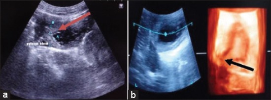
Results: Present study was done to compare 2D and 3D ultrasonographic images in different gastric affections in dogs. In case of uremic gastropathy due to inflammatory response, the wall of the stomach was 0.6 cm thick and hyperechoic and gastric folds were also hyperechoic indicative of gastritis. In second, third, and fourth case of gastritis the wall of the stomach was 0.7, 0.6, and 0.55 cm, respectively thick and hyperechoic. In fifth and sixth case of gastritis, inflammatory response due to ingestion of polythene and sand led to gastritis and ultrasonographically, the wall of the stomach was 0.6 cm and 0.7 cm thick, respectively, and hyperechoic. In case of gastric ulcer, ultrasonographically, there was a disruption of gastric mucosal layer. In cases of gastric dilatation, anechoic content indicating fluid was seen in stomach area and due to dilatation boundary of the stomach was not clear and the increase in the lumen of the stomach was observed. In case of foreign body, ultrasonographically the wall of the stomach was 0.55 cm thick and hyperechoic. In the middle of the stomach, multiple hyperechoic shadows of the foreign bodies i.e. leather and bunch of straw of grass were observed. In case of pyloric stenosis ultrasonographically, anechoic lumen of the pylorus surrounded by 0.5 cm hypoechoic thickened muscle. In some cases, 3D ultrasonography was not diagnostic i.e. gastric foreign bodies and gastric dilatation. These conditions were better visualized on the 2D sonogram.
Conclusion: The appearance of clinical conditions of the stomach such as gastritis and pyloric stenosis were more distinct on 3D ultrasonogram than 2D ultrasonogram. The 3D ultrasonography was not diagnostic in cases of gastric foreign bodies and gastric dilatation.
Keywords: dogs; gastric dilatation; gastric foreign bodies; gastric ulcer; gastritis; three-dimensional ultrasonogram.
Figures


Similar articles
-
Diagnosis of urinary bladder diseases in dogs by using two-dimensional and three-dimensional ultrasonography.Vet World. 2015 Jul;8(7):819-22. doi: 10.14202/vetworld.2015.819-822. Epub 2015 Jul 7. Vet World. 2015. PMID: 27047159 Free PMC article.
-
Diagnosis of pathological conditions of kidney by two-dimensional and three-dimensional ultrasonographic imaging in dogs.Vet World. 2016 Jul;9(7):693-8. doi: 10.14202/vetworld.2016.693-698. Epub 2016 Jul 4. Vet World. 2016. PMID: 27536028 Free PMC article.
-
Double contrast-enhanced two-dimensional and three-dimensional ultrasonography for evaluation of gastric lesions.World J Gastroenterol. 2012 Aug 21;18(31):4136-44. doi: 10.3748/wjg.v18.i31.4136. World J Gastroenterol. 2012. PMID: 22919245 Free PMC article.
-
Application of Ultrasound in Detecting and Removing Migrating Grass Awns in Dogs and Cats: A Systematic Review.Animals (Basel). 2023 Jun 22;13(13):2071. doi: 10.3390/ani13132071. Animals (Basel). 2023. PMID: 37443870 Free PMC article. Review.
-
The Morbid Anatomy of the Bowels, Liver, and Stomach, Illustrated by a Series of Plates, and a Summary of the Symptoms of the Acute and Chronic Affections of the Above-Named Organs.Med Chir Rev. 1828 Oct 1;9(18):347-358. Med Chir Rev. 1828. PMID: 29917621 Free PMC article. Review. No abstract available.
References
-
- Gomaa M, Samy M.T, Omar M.S.A, Mekkawy N.H. Ultrasonographic findings of most common surgical disorders of gastrointestinal tract in dogs and cats. Iran. J. Vet. Surg. 2012;7(1):23–28.
-
- Gilja O.H. Ultrasound of the stomach. Ultraschall, Med. 2007;28(1):32–39. - PubMed
-
- Larson M.M, Biller D.S. Ultrasound of gastrointestinal tract. Vet. Clin. Small Anim. 2009;39:747–759. - PubMed
-
- Trahair L, Jones K.L. Ultrasonography of the stomach. Applied Aspects of Ultrasonography in Humans. InTech, Rijeka, Croatia. 2012:175–190.
-
- Choi M, Seo M, Jung J, Lee K, Yoon J, Chang D, Park R.D. Evaluation of canine gastric motility with Ultrasonography. J. Vet. Med. Sci. 2002;64(1):17–21. - PubMed
LinkOut - more resources
Full Text Sources
Other Literature Sources