. 2016 Mar 31:10:109.
doi: 10.3389/fnins.2016.00109. eCollection 2016.

Electrophysiology of Hypothalamic Magnocellular Neurons In vitro: A Rhythmic Drive in Organotypic Cultures and Acute Slices

Affiliations

Electrophysiology of Hypothalamic Magnocellular Neurons In vitro: A Rhythmic Drive in Organotypic Cultures and Acute Slices

Jean-Marc Israel et al. Front Neurosci. .

Abstract

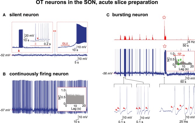
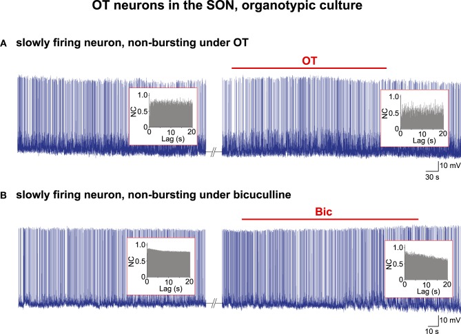
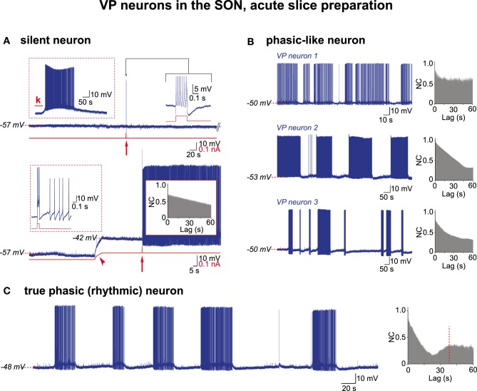
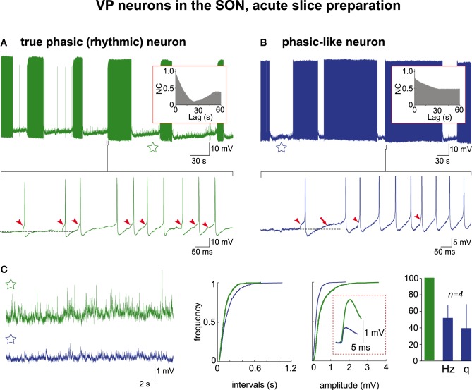
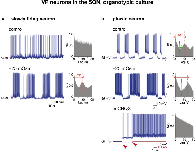
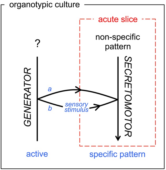
Hypothalamic neurohormones are released in a pulsatile manner. The mechanisms of this pulsatility remain poorly understood and several hypotheses are available, depending upon the neuroendocrine system considered. Among these systems, hypothalamo-neurohypophyseal magnocellular neurons have been early-considered models, as they typically display an electrical activity consisting of bursts of action potentials that is optimal for the release of boluses of the neurohormones oxytocin and vasopressin. The cellular mechanisms underlying this bursting behavior have been studied in vitro, using either acute slices of the adult hypothalamus, or organotypic cultures of neonatal hypothalamic tissue. We have recently proposed, from experiments in organotypic cultures, that specific central pattern generator networks, upstream of magnocellular neurons, determine their bursting activity. Here, we have tested whether a similar hypothesis can be derived from in vitro experiments in acute slices of the adult hypothalamus. To this aim we have screened our electrophysiological recordings of the magnocellular neurons, previously obtained from acute slices, with an analysis of autocorrelation of action potentials to detect a rhythmic drive as we recently did for organotypic cultures. This confirmed that the bursting behavior of magnocellular neurons is governed by central pattern generator networks whose rhythmic drive, and thus probably integrity, is however less satisfactorily preserved in the acute slices from adult brains.

Keywords: burst firing; oxytocin; pulse generator; supraoptic nucleus; vasopressin.


Figures

Figure 1
Figure 1
Electrical activity in OT neurons in hypothalamic acute slices. (A) A silent OT cell displaying rare action potentials (APs, asterisks) was subjected to two depolarizing challenges (insets). Left inset, a brief positive current pulse (50 ms, 0.1 nA; red trace; expanded from the large arrowhead) triggered a 10 s-long decaying burst of APs (the small arrowhead shows the DAP maintaining firing). Right inset, glutamate (GLU) in the perfusion medium (10−5 M, 30 s) induced a strong depolarization triggering a sustained firing. (B) A continuously active OT neuron with irregular firing with no rhythmic drive from the APs autocorrelation analysis (inset). (C) Exceptional example of a truly bursting OT neuron. Top, rate-meter showing the high-frequency burst (HFB) of action potentials (star). Middle, respective raw recording and associated autocorrelogram (inset) showing rhythmic drive. Bottom, expanded traces showing the importance of the EPSPs (arrowheads) in initiating the burst (left) and HFB (right) and triggering the APs within the burst (middle) (note that APs were truncated in the right trace).
Figure 2
Figure 2
Electrical activity in OT neurons in organotypic cultures. Examples of two spontaneously firing cells that did not change their pattern of activity under bath application of (A) the OT peptide (10−5 M) or (B) the GABAA receptor antagonist bicuculline (Bic; 10−5 M). No rhythmic drive was detected by the AAA (insets) either before or during the application of the burst-inducing agents.
Figure 3
Figure 3
Electrical activities in VP neurons in hypothalamic acute slices. (A) A silent VP cell displaying action potentials only if subjected to depolarizing challenges. Top trace, left inset, sustained firing in response to kainate (k) in perfusion medium (10−5 M; 30 s); right inset, a train of APs (expanded trace; truncated) in response to a brief positive current pulse (0.2 s, 0.15 nA; red arrow). Bottom trace, the same neuron when depolarized by constant current injection (red trace, arrowhead) now displays a prolonged firing in response to a brief current pulse (0.1 s, 0.1 nA) (left inset, expanded from arrow). Note absence of rhythmic drive (right inset). (B) Three examples of phasic-like activities displayed by VP neurons. Neuron 1, bursts and silent periods of irregular duration. Neuron 2, bursts and silent periods of similar duration. Neuron 3, short and long bursts, irregular silent periods. Autocorrelograms show no rhythmic drive. (C) A truly phasic activity sustained by a rhythmic drive.
Figure 4
Figure 4
Electrical activities in VP neurons in hypothalamic acute slices. Mechanisms of burst generation in spontaneously active neurons (A) with and (B) without rhythmic drive (autocorrelograms in the insets). The raw recordings (upper traces) are expanded (lower traces) to reveal that the first AP in the burst is triggered by an EPSP (arrowheads) and is followed by a DAP (arrow) in (B) only (APs are truncated in B). The subsequent APs in the burst are essentially triggered by EPSPS in (A) and by both EPSPs and DAPs in (B). Note in the samples of inter-burst activity (stars) magnified in (C) the heightened synaptic activity in (A) reflected by the cumulative frequencies of EPSPs' intervals and amplitudes (mean amplitudes in dashed frame). The histogram shows that phasic-like neurons display EPSPs at lower frequency (Hz) and smaller amplitude (q) (green column = 100% for both frequency and amplitude of events recorded in truly rhythmic neurons; n = 4 each).
Figure 5
Figure 5
Electrical activities in VP neurons in hypothalamic organotypic cultures. (A) Transition from irregular (top, control medium) to phasic (bottom, +25 mOsm medium) activity in a VP neuron by a hyperosmotic stimulus. Note rhythmic drive [with a cycling period (c.p.) of 28 s] in phasic firing-mode only. (B) Increase in rhythm quality in a spontaneously phasic VP neuron (top, control medium) by a hyperosmotic stimulus (middle, +25 mOsm medium) and the dependence of phasic firing on glutamatergic inputs (bottom). Note that hyperosmolality increases the cycling period (c.p.) and the quality of the rhythm (a), while CNQX (10−6 M) abolishes spiking and rhythmic drive (depolarization by two successive current injections (arrowheads) induces spontaneous firing not supported by a rhythmic drive).
Figure 6
Figure 6
Schematic diagram of functional state transition of the secretomotor OT and VP neurons in vitro. As visible in our acute slice model (dashed red frame) and in our organotypic culture model (black frame), the transition from random firing to the typical neurosecretory bursting/phasic pattern is either inherent (a, developmentally active: OT neurons) or gated (b, elicited by sensory stimuli: VP neurons). The acute slice model in most cases (99% for OT neurons, 70% for VP neurons) does not include the generator network. See Conclusion.
Figure 7
Figure 7
Analogy between the locomotor and neuroendocrine circuits. Motor neurons and neuroendocrine cells are output units driven by central pattern generator (CPG) networks. CPGs generate the specific electrical firing required for secretion of neurotransmitters/neurohormones and the desired action of the effector structure: skeletal muscle for alpha-motor neurons, uterus and mammary myoepithelial cells in the case of magnocellular oxytocin (OT) neurons. The presumptive “OT-CPG” necessary for the milk-ejection reflex may be a two-level CPG comprising a rhythmogenic component in interaction with a pattern-forming component, the output of which being bursting activity. The secretomotor unit is mainly a follower of the CPG output triggering neurosecretion. BBB, blood-brain-barrier. For details, see the Conclusion.

Similar articles

Cited by

References

    1. Andrew R. D., Dudek F. E. (1983). Burst discharge in mammalian neuroendocrine cells involves an intrinsic regenerative mechanism. Science 221, 1050–1052. 10.1126/science.6879204 - DOI - PubMed
    1. Armstrong W. E., Smith B. N., Tian M. (1994). Electrophysiological characteristics of immunochemically identified rat oxytocin and vasopressin neurones in vitro. J. Physiol. 475, 115–128. 10.1113/jphysiol.1994.sp020053 - DOI - PMC - PubMed
    1. Armstrong W. E., Stern J. E. (1998). Electrophysiological distinctions between oxytocin and vasopressin neurons in the supraoptic nucleus. Adv. Exp. Med. Biol. 449, 67–77. 10.1007/978-1-4615-4871-3_7 - DOI - PubMed
    1. Armstrong W. E., Wang L., Li C., Teruyama R. (2010). Performance, properties and plasticity of identified oxytocin and vasopressin neurones in vitro. J. Neuroendocrinol. 22, 330–342. 10.1111/j.1365-2826.2010.01989.x - DOI - PMC - PubMed
    1. Ben-Barak Y., Russell J. T., Whitnall M. H., Ozato K., Gainer H. (1985). Neurophysin in the hypothalamo-neurohypophysial system: I. production and characterization of monoclonal antibodies. J. Neurosci. 5, 81–97. - PMC - PubMed

LinkOut - more resources