

EEG Resting State Functional Connectivity Analysis in Children with Benign Epilepsy with Centrotemporal Spikes
- PMID: 27065797
- PMCID: PMC4815534
- DOI: 10.3389/fnins.2016.00143
EEG Resting State Functional Connectivity Analysis in Children with Benign Epilepsy with Centrotemporal Spikes
Abstract
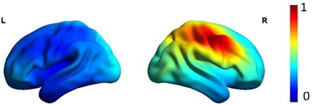
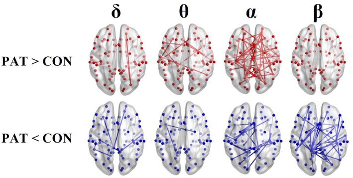
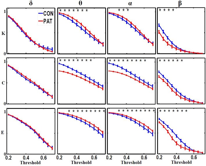
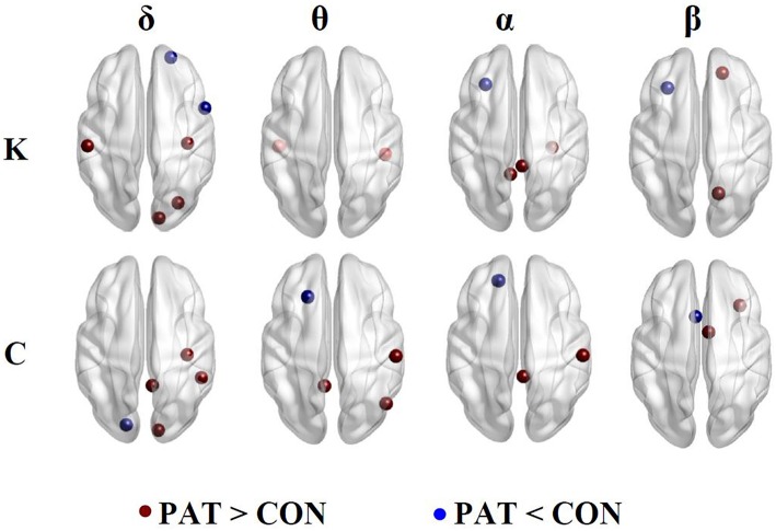
In this study, we investigated changes in functional connectivity (FC) of the brain networks in patients with benign epilepsy with centrotemporal spikes (BECTS) compared to healthy controls using high-density EEG data collected under eyes-closed resting state condition. EEG source reconstruction was performed with exact Low Resolution Electromagnetic Tomography (eLORETA). We investigated FC between 84 Brodmann areas using lagged phase synchronization (LPS) in four frequency bands (δ, θ, α, and β). We further computed the network degree, clustering coefficient and efficiency. Compared to controls, patients displayed higher θ and α and lower β LPS values. In these frequency bands, patients were also characterized by less well ordered brain networks exhibiting higher global degrees and efficiencies and lower clustering coefficients. In the β band, patients exhibited reduced functional segregation and integration due to loss of both local and long-distance functional connections. These findings suggest that benign epileptic brain networks might be functionally disrupted due to their altered functional organization especially in the α and β frequency bands.
Keywords: centrotemporal spikes; children epilepsy; functional connectivity; graph theory; phase synchronization; resting sate.
Figures
Similar articles
-
Electroencephalographic Resting-State Functional Connectivity of Benign Epilepsy with Centrotemporal Spikes.J Clin Neurol. 2019 Apr;15(2):211-220. doi: 10.3988/jcn.2019.15.2.211. J Clin Neurol. 2019. PMID: 30938108 Free PMC article.
-
EEG resting state analysis of cortical sources in patients with benign epilepsy with centrotemporal spikes.Neuroimage Clin. 2015 Sep 3;9:275-82. doi: 10.1016/j.nicl.2015.08.014. eCollection 2015. Neuroimage Clin. 2015. PMID: 26509114 Free PMC article.
-
Altered brain functional connectivity induced by physical exercise may improve neuropsychological functions in patients with benign epilepsy.Epilepsy Behav. 2017 Nov;76:126-132. doi: 10.1016/j.yebeh.2017.06.021. Epub 2017 Sep 15. Epilepsy Behav. 2017. PMID: 28919388
-
Resting-State EEG Functional Connectivity in Children with Rolandic Spikes with or without Clinical Seizures.Biomedicines. 2022 Jun 29;10(7):1553. doi: 10.3390/biomedicines10071553. Biomedicines. 2022. PMID: 35884857 Free PMC article.
-
Functional and Structural Network Disorganizations in Typical Epilepsy With Centro-Temporal Spikes and Impact on Cognitive Neurodevelopment.Front Neurol. 2019 Aug 29;10:809. doi: 10.3389/fneur.2019.00809. eCollection 2019. Front Neurol. 2019. PMID: 31555191 Free PMC article. Review.
Cited by
-
Spectral-power associations reflect amplitude modulation and within-frequency interactions on the sub-second timescale and cross-frequency interactions on the seconds timescale.PLoS One. 2020 May 18;15(5):e0228365. doi: 10.1371/journal.pone.0228365. eCollection 2020. PLoS One. 2020. PMID: 32421714 Free PMC article.
-
Characterisation of ictal and interictal states of epilepsy: A system dynamic approach of principal dynamic modes analysis.PLoS One. 2018 Jan 19;13(1):e0191392. doi: 10.1371/journal.pone.0191392. eCollection 2018. PLoS One. 2018. PMID: 29351559 Free PMC article.
-
Spatiotemporal dynamics of maximal and minimal EEG spectral power.PLoS One. 2021 Jul 20;16(7):e0253813. doi: 10.1371/journal.pone.0253813. eCollection 2021. PLoS One. 2021. PMID: 34283869 Free PMC article.
-
Effect of interictal epileptiform discharges on EEG-based functional connectivity networks.Clin Neurophysiol. 2020 May;131(5):1087-1098. doi: 10.1016/j.clinph.2020.02.014. Epub 2020 Mar 4. Clin Neurophysiol. 2020. PMID: 32199397 Free PMC article.
-
Altered brain network topology related to working memory in internet addiction.J Behav Addict. 2020 Jul 7;9(2):325-338. doi: 10.1556/2006.2020.00020. Print 2020 Jun. J Behav Addict. 2020. PMID: 32644933 Free PMC article.
References
-
- Adebimpe A. A., Aarabi A., Bourel E., Mahmoudzadeh M., Wallois F. (2016). ID 234 – brain network analysis of EEG functional connectivity in patients with benign childhood epilepsy with centro-temporal spikes. Clin. Neurophysiol. 127:e60 10.1016/j.clinph.2015.11.198 - DOI
-
- Adebimpe A. A., Aarabi A., Mahmoudzadeh M., Bourel-Ponchel E., Wallois F. (2014). LP7: the comparison of resting state networks between normal children and adolescents with benign childhood epilepsy with centrotemporal spikes: a high density EEG study. Clin. Neurophysiol. 125, S80–S81. 10.1016/s1388-2457(14)50268-4 - DOI
LinkOut - more resources
Full Text Sources
Other Literature Sources