. 2016 Mar 29:10:77.
doi: 10.3389/fncel.2016.00077. eCollection 2016.

Estrogen Receptor Beta and 2-arachidonoylglycerol Mediate the Suppressive Effects of Estradiol on Frequency of Postsynaptic Currents in Gonadotropin-Releasing Hormone Neurons of Metestrous Mice: An Acute Slice Electrophysiological Study

Affiliations

Estrogen Receptor Beta and 2-arachidonoylglycerol Mediate the Suppressive Effects of Estradiol on Frequency of Postsynaptic Currents in Gonadotropin-Releasing Hormone Neurons of Metestrous Mice: An Acute Slice Electrophysiological Study

Flóra Bálint et al. Front Cell Neurosci. .

Abstract

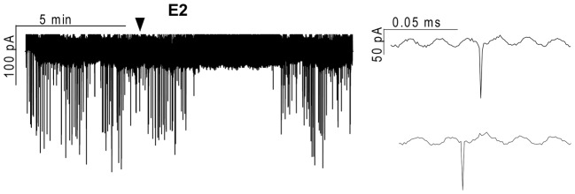
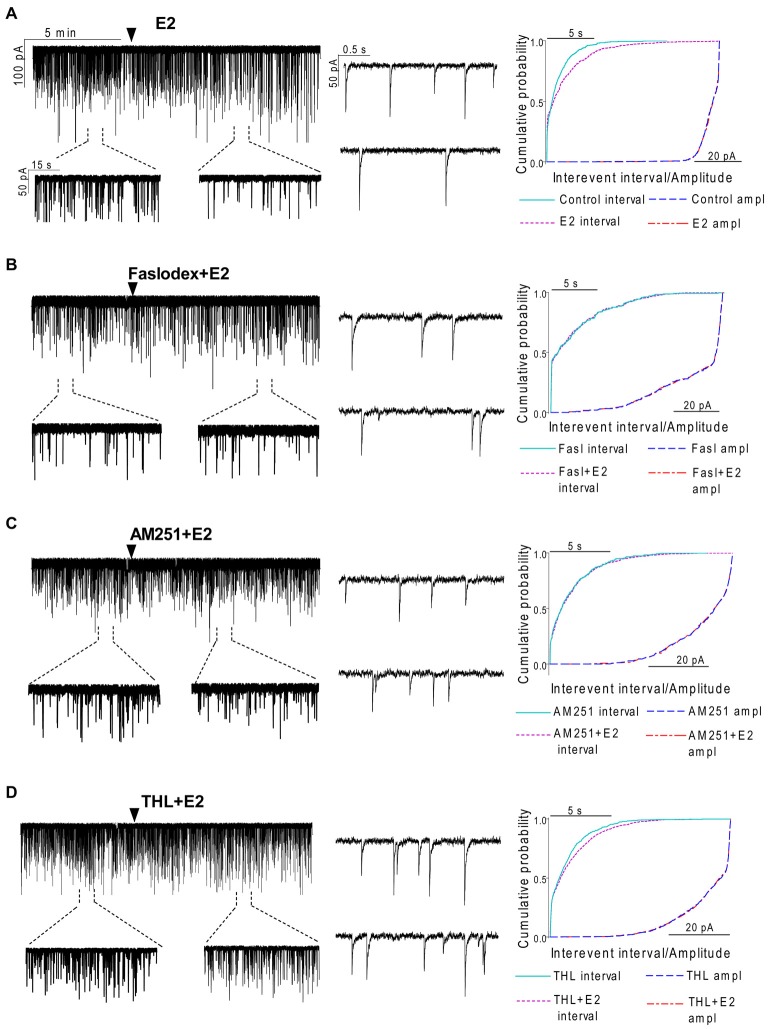
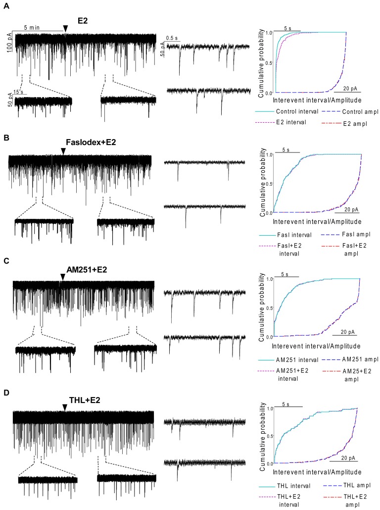
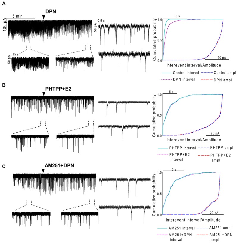
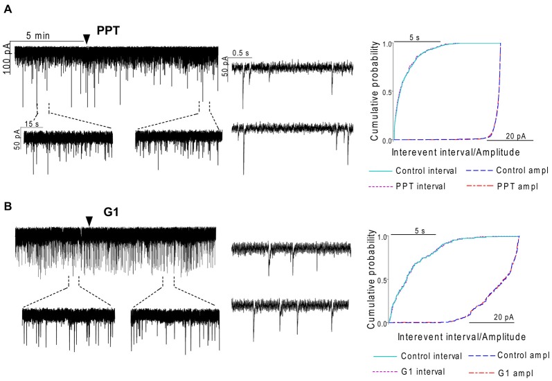
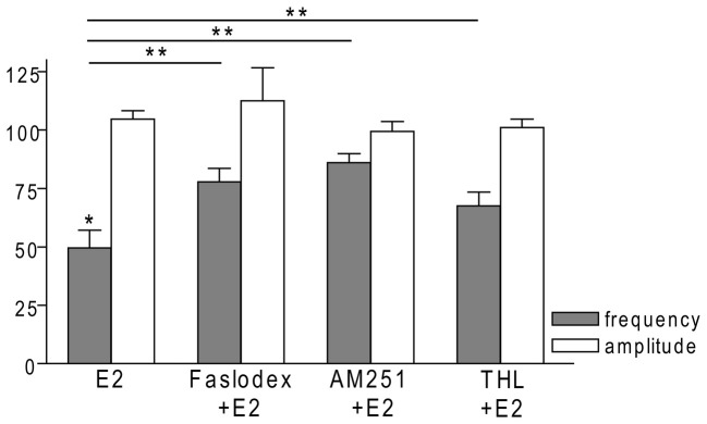
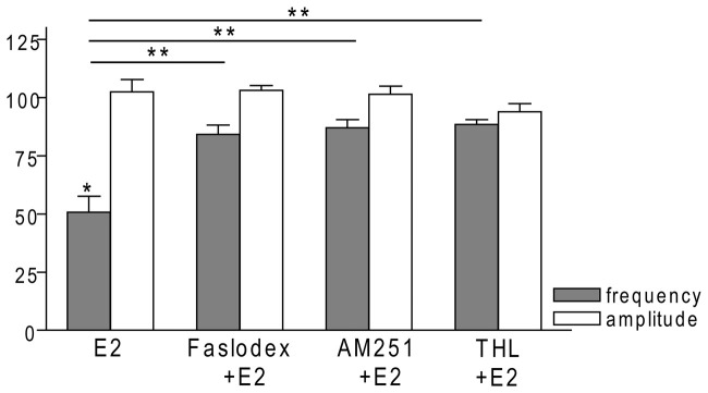
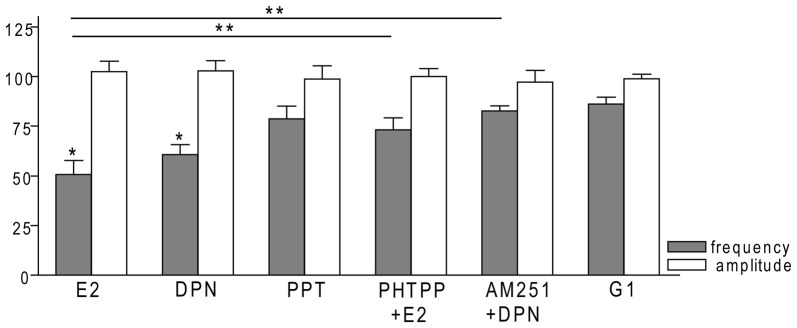
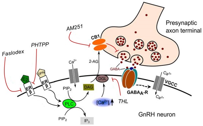
Gonadotropin-releasing hormone (GnRH) neurons are controlled by 17β-estradiol (E2) contributing to the steroid feedback regulation of the reproductive axis. In rodents, E2 exerts a negative feedback effect upon GnRH neurons throughout the estrus-diestrus phase of the ovarian cycle. The present study was undertaken to reveal the role of estrogen receptor subtypes in the mediation of the E2 signal and elucidate the downstream molecular machinery of suppression. The effect of E2 administration at low physiological concentration (10 pM) on GnRH neurons in acute brain slices obtained from metestrous GnRH-green fluorescent protein (GFP) mice was studied under paradigms of blocking or activating estrogen receptor subtypes and interfering with retrograde 2-arachidonoylglycerol (2-AG) signaling. Whole-cell patch clamp recordings revealed that E2 significantly diminished the frequency of spontaneous postsynaptic currents (sPSCs) in GnRH neurons (49.62 ± 7.6%) which effect was abolished by application of the estrogen receptor (ER) α/β blocker Faslodex (1 μM). Pretreatment of the brain slices with cannabinoid receptor type 1 (CB1) inverse agonist AM251 (1 μM) and intracellularly applied endocannabinoid synthesis blocker THL (10 μM) significantly attenuated the effect of E2 on the sPSCs. E2 remained effective in the presence of tetrodotoxin (TTX) indicating a direct action of E2 on GnRH cells. The ERβ specific agonist DPN (10 pM) also significantly decreased the frequency of miniature postsynaptic currents (mPSCs) in GnRH neurons. In addition, the suppressive effect of E2 was completely blocked by the selective ERβ antagonist PHTPP (1 μM) indicating that ERβ is required for the observed rapid effect of the E2. In contrast, the ERα agonist PPT (10 pM) or the membrane-associated G protein-coupled estrogen receptor (GPR30) agonist G1 (10 pM) had no significant effect on the frequency of mPSCs in these neurons. AM251 and tetrahydrolipstatin (THL) significantly abolished the effect of E2 whereas AM251 eliminated the action of DPN on the mPSCs. These data suggest the involvement of the retrograde endocannabinoid mechanism in the rapid direct effect of E2. These results collectively indicate that estrogen receptor beta and 2-AG/CB1 signaling mechanisms are coupled and play an important role in the mediation of the negative estradiol feedback on GnRH neurons in acute slice preparation obtained from intact, metestrous mice.

Keywords: 2-AG; CB1; GABA; GnRH neuron; estrogen receptor beta; negative estrogen feedback; retrograde signaling.


Figures

Figure 1
Figure 1
Effect of 17β-estradiol (E2) on the action currents of gonadotropin-releasing hormone (GnRH) neurons in brain slice of metestrous female mouse. Application of 10 pM E2 resulted in a significant decrease in the frequency of the action currents on GnRH neurons. Individual events during the control phase (upper inset) and the E2-treated phase (lower inset) present no change. Arrowhead shows the onset of E2 administration.
Figure 2
Figure 2
Effect of E2 on the spontaneous postsynaptic currents (sPSCs) of GnRH neurons in brain slice of metestrous female mice. (A) E2 in low concentration (10 pM) decreased the frequency of the sPSCs with no change in the average amplitude of them. One-minute-long periods of the recording before and after application of E2 are depicted under the recording. Cumulative probability plot presents reduction in the interevent intervals, but not in amplitude. (B) Pretreatment of the brain slice with the non-selective ER antagonist Faslodex (1 μM, 10 min) inhibited the effect of E2 on the sPSCs. (C) Effect of E2 on the sPSCs was abolished by the pretreatment with cannabinoid receptor type 1 (CB1) inverse agonist AM251 (1 μM, 10 min). (D) Similar inhibition was observed in case of intracellularly applied diacylglycerol (DAG) lipase inhibitor THL (10 μM, 20 min). Individual events of sPSCs show no change in waveform properties in the treated phase (lower insets) as compared to the control phase (upper insets) in all sPSC measurements. Cumulative probability plots in (B–D) graphs present no change in interevent intervals and amplitudes. Arrowhead shows the onset of drug administration.
Figure 3
Figure 3
Effect of E2 on the miniature postsynaptic currents (mPSCs) of GnRH neurons in the presence of non-selective estrogen receptor (ER) antagonist and endocannabinoid receptor/synthesis blockers. (A) E2 decreased the frequency of the mPSCs with no change in the average amplitude of them. One-minute-long periods of the recording before and after application of E2 are illustrated under the recording. Cumulative probability plot presents reduction in the interevent intervals, but not in amplitude in the case of the effect of E2 alone on the mPSCs. (B) Pretreatment of the brain slice with the non-selective ER antagonist Faslodex (1 μM, 10 min) inhibited the action of E2 on the mPSCs. (C) Effect of E2 on the mPSCs was abolished by pretreatment with cannabinoid receptor type 1 (CB1) inverse agonist AM251 (1 μM, 10 min). (D) Similar inhibition was observed in case of intracellularly applied DAG lipase inhibitor THL (10 μM, 20 min). Arrowhead shows the onset of drug administration. Individual events of mPSC in the control phase (upper insets) and the treated phase (lower insets) show no change in waveform properties in any measurements (A—D). Cumulative probability plots of treatments in (B–D) graphs show no change in interevent intervals or amplitudes.
Figure 4
Figure 4
The effect of ERβ activation on the mPSCs in GnRH neurons of the metestrous female mice: outcome of ERβ antagonist, agonist and CB1 blocker. (A) The subtype-selective ERβ agonist DPN (10 pM, 10 min) significantly decreased the frequency of mPSCs. One-minute long periods of the recording before and after application of the agonist are illustrated under the recording. Cumulative probability plot presents reduction of the interevent intervals, but not in amplitude. (B) Pretreatment of the brain slice with the selective ERβ receptor antagonist PHTPP (1 μM, 10 min) inhibited the effect of E2 on mPSCs. (C) ERβ agonist DPN had no significant effect on the frequency of mPSCs in the presence of AM251 (1 μM, 10 min). Arrowhead shows the onset of drug administration. Individual events of mPSC show no change in waveform properties of the treated phase (lower insets) as compared to the control phase (upper insets) in any of the mPSC measurements. Cumulative probability plots show no change in interevent intervals and amplitudes in (B,C) graphs.
Figure 5
Figure 5
The selective ERα agonist and G protein-coupled estrogen receptor (GPR30) agonist exert no effects on the mPSCs in GnRH neurons of the metestrous female mice. (A) The selective ERα agonist PPT (10 pM, 10 min) was unable to modify the frequency of mPSCs in the recorded GnRH neurons. (B) Similarly, the GPR30 receptor agonist G1 (10 pM; 10 min) did not modify the frequency of the mPSCs. Arrowhead shows the onset of drug administration. Individual events of mPSC show no change in waveform properties of the treated phase (lower insets) compared to the control phase (upper insets) in each mPSC measurements. Cumulative probability plots of the treatments show no change in interevent intervals or amplitudes.
Figure 6
Figure 6
Bar graph summarizing the percentage changes in the frequency and the amplitude of the sPSCs resulting from E2 treatment in the presence of Faslodex, AM251 and THL. E2 significantly decreased the frequency of sPSCs. Inhibition of its effect could be achieved with antagonizing the ERs, CB1 receptors or blocking the intracellular 2-AG endocannabinoid synthesis. The amplitude of the mPSCs did not change in any of the treatments. *p < 0.05 as compared to the control; **p < 0.05 as compared to the change evoked by E2 treatment.
Figure 7
Figure 7
Bar graph summarizing the percentage changes in the frequency and the amplitude of the mPSCs resulting from E2 treatment in the presence of Faslodex, AM251 and THL. E2 significantly decreased the frequency of mPSCs. Inhibition of this effect could be achieved with antagonizing the ERs by Faslodex. Effect of E2 was eliminated by the pretreatment with CB1 inverse agonist AM251 or the intracellularly applied 2-AG endocannabinoid synthesis blocker THL. The amplitude of the mPSCs did not change in any of the treatments. *p < 0.05 as compared to the control; **p < 0.05 as compared to the change evoked by E2 treatment.
Figure 8
Figure 8
Bar graph summarizing the percentage changes in the frequency and the amplitude of the mPSCs resulting from selective ER agonists and various antagonists. The E2 and the selective ERβ agonist DPN significantly decreased the frequency of mPSCs. Effect of DPN was eliminated by the pretreatment with CB1 inverse agonist AM251. Effect of E2 could be inhibited by antagonizing selectively the ERβ by PHTPP. The selective ERα agonist PPT and the GPR30 receptor agonist G1 had no significant effect on the frequency of mPSCs. The amplitude of the mPSCs presented no change in any of the treatments. *p < 0.05 compare to the control; **p < 0.05 as compared to the change caused by E2 treatment.
Figure 9
Figure 9
Schematic illustration of the interaction between E2 and endocannabinoid signaling in GnRH neuron of the metestrous female mice. Binding of E2 to ERβ activates synthesis and release of 2-AG in the GnRH neuron. The released endocannabinoid 2-AG then binds to CB1 expressed in the presynaptic terminal of GABAergic afferents and causes suppression of GABA release into the synaptic cleft. This effect of E2 was blocked when the non-selective ER antagonist (Faslodex) or the selective ERβ receptor antagonist (PHTPP) was administered. The signaling was also inhibited when the CB1 inverse agonist (AM251) or the DAG lipase inhibitor (THL) was applied. E2, 17β-estradiol; ERβ, estrogen receptor beta; DPN, subtype selective ERβ agonist; DAG, diacylglycerol; DGL, DAG-lipase; CB1, cannabinoid receptor type-1; AM251, CB1 inverse agonist; Faslodex, non-selective estrogen receptor antagonist; PHTPP, subtype selective ERβ antagonist; 2-AG, 2-arachidonoylglycerol; THL, tetrahydrolipstatin (DAG-lipase inhibitor); PIP2, phosphatidylinositol 4,5-bisphosphate; IP3, inositol 1,4,5-trisphosphate; PLC, phospholipase-C; GABAA-R, GABAA receptor; [CaI2+], intracellular free calcium; VGCC, voltage-gated calcium channel. Dashed arrow denote putative action.

References

    1. Abe H., Keen K. L., Terasawa E. (2008). Rapid action of estrogens on intracellular calcium oscillations in primate luteinizing hormone-releasing hormone-1 neurons. Endocrinology 149, 1155–1162. 10.1210/en.2007-0942 - DOI - PMC - PubMed
    1. Abe H., Terasawa E. (2005). Firing pattern and rapid modulation of activity by estrogen in primate luteinizing hormone releasing hormone-1 neurons. Endocrinology 146, 4312–4320. 10.1210/en.2005-0435 - DOI - PMC - PubMed
    1. Abrahám I. M., Han S. K., Todman M. G., Korach K. S., Herbison A. E. (2003). Estrogen receptor beta mediates rapid estrogen actions on gonadotropin-releasing hormone neurons in vivo. J. Neurosci. 23, 5771–5777. - PMC - PubMed
    1. Attardi B., Hotchkiss J., Knobil E. (1980). Monkey pituitary oestrogen receptors and the biphasic action of oestradiol on gonadotropin secretion. Nature 285, 252–254. 10.1038/285252a0 - DOI - PubMed
    1. Byers S. L., Wiles M. V., Dunn S. L., Taft R. A. (2012). Mouse estrous cycle identification tool and images. PLoS One 7:e35538. 10.1371/journal.pone.0035538 - DOI - PMC - PubMed

LinkOut - more resources