Review
. 2016 Mar 31:7:119.
doi: 10.3389/fphys.2016.00119. eCollection 2016.

Insulin Is a Key Modulator of Fetoplacental Endothelium Metabolic Disturbances in Gestational Diabetes Mellitus

Affiliations
Review

Insulin Is a Key Modulator of Fetoplacental Endothelium Metabolic Disturbances in Gestational Diabetes Mellitus

Luis Sobrevia et al. Front Physiol. .

Abstract

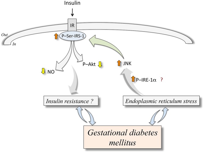
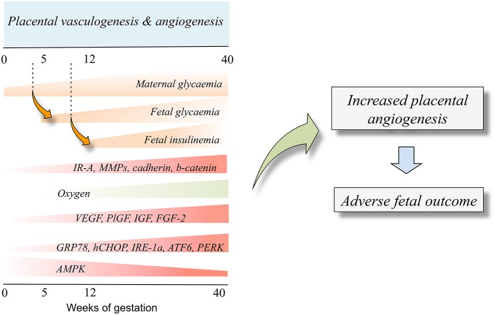
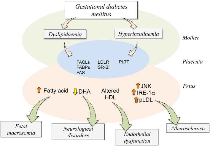
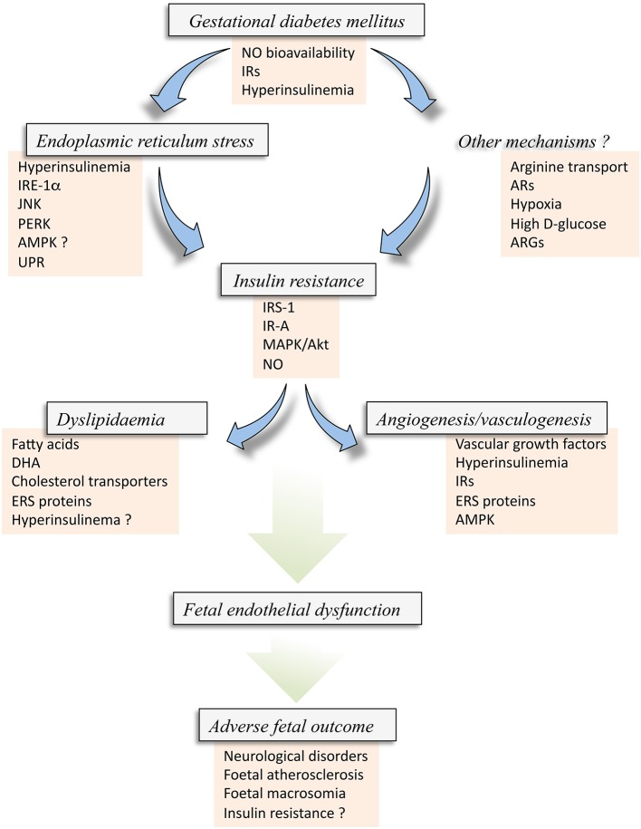
Gestational diabetes mellitus (GDM) is a disease of the mother that associates with altered fetoplacental vascular function. GDM-associated maternal hyperglycaemia result in fetal hyperglycaemia, a condition that leads to fetal hyperinsulinemia and altered L-arginine transport and synthesis of nitric oxide, i.e., endothelial dysfunction. These alterations in the fetoplacental endothelial function are present in women with GDM that were under diet or insulin therapy. Since these women and their newborn show normal glycaemia at term, other factors or conditions could be altered and/or not resolved by restoring normal level of circulating D-glucose. GDM associates with metabolic disturbances, such as abnormal handling of the locally released vasodilator adenosine, and biosynthesis and metabolism of cholesterol lipoproteins, or metabolic diseases resulting in endoplasmic reticulum stress and altered angiogenesis. Insulin acts as a potent modulator of all these phenomena under normal conditions as reported in primary cultures of cells obtained from the human placenta; however, GDM and the role of insulin regarding these alterations in this disease are poorly understood. This review focuses on the potential link between insulin and endoplasmic reticulum stress, hypercholesterolemia, and angiogenesis in GDM in the human fetoplacental vasculature. Based in reports in primary culture placental endothelium we propose that insulin is a factor restoring endothelial function in GDM by reversing ERS, hypercholesterolaemia and angiogenesis to a physiological state involving insulin activation of insulin receptor isoforms and adenosine receptors and metabolism in the human placenta from GDM pregnancies.

Keywords: angiogenesis; endoplasmic reticulum stress; endothelium; gestational diabetes; insulin; lipids; placenta.


Figures

Figure 1
Figure 1
Endoplasmic reticulum stress and abnormal insulin signaling in human fetoplacental endothelium from gestational diabetes mellitus. Gestational diabetes mellitus is a disease that associates with endoplasmic reticulum stress (ERS). The latter is an abnormal metabolic condition that could (?) lead to increased (⇧) phosphorylation of inositol-requiring enzyme 1α (P-IRE-1α) resulting in higher c-Jun N-terminal kinase (JNK) activity. This phenomenon causes phosphorylation of insulin receptor substrate 1 at serine307 (P-Ser-IRS-1) ending in lower insulin receptor (IR)-associated cell signaling in response to insulin. This altered response to insulin results in reduced (⇩) synthesis of nitric oxide (NO) and phosphorylation of protein kinase B/Akt (P-Akt). These mechanisms are proposed to be potentially involved in insulin resistance (Insulin resistance?) in the human fetoplacental endothelium. At present, it is unclear whether GDM causes insulin resistance or ERS, or are these abnormal metabolic conditions that result in GDM clinical manifestations. Composed from information reported by Ozcan et al. (2004, 2006), Taniguchi et al. (2006), Eizirik et al. (2008), Hotamisligil (2010), Sáez et al. (2014), Westermeier et al. (2011, 2015a).
Figure 2
Figure 2
Fetal insulinemia and altered angiogenesis in fetoplacental endothelium from gestational diabetes mellitus. With the progression of pregnancy up to the 40th weeks of gestation, the maternal glycaemia increases, and could reach supraphysiological levels in pregnancies where the mother is diagnosed with gestational diabetes mellitus. The maternal hyperglycaemia results in increased fetal glycaemia from about the 5th week of gestation (dotted line), a condition resulting in a supraphysiological increase of fetal insulinemia from the 12th week of gestation. Increased fetal insulinemia results in altered placental vascular development and growth leading to angiogenesis alterations (Placental vasculogenesis and angiogenesis). Thus, an adverse fetal outcome is seen as a result of abnormal angiogenesis. Cell signaling mechanisms involved in this phenomenon include altered expression and/or activity of several molecules that are responsive to insulin (IR-A, MMPs, cadherin, b-catenin). Equally, a low oxygen level at the beginning of pregnancy increases the expression of proangiogenic growth factors (VEGF, PlGF, IGF, FGF-2) and increased (GRP78, CHOP, IRE-1α, ATF6, PERK) or reduced (AMPK) expression and/or activity. Composed from information reported by Babawale et al. (2000), Jirkovská et al. (2002), Easwaran et al. (2003), Baumüller et al. (2015), Westermeier et al. (2015b).
Figure 3
Figure 3
Potential consequences of dyslipidaemia and hyperinsulinemia on the human fetoplacental unit from gestational diabetes mellitus. Gestational diabetes mellitus (GDM) results in maternal (Mother) metabolic alterations leading to dyslipidaemia and hyperinsulinemia. These two abnormal metabolic conditions are associated with higher expression and activity of several molecules involved in the placental transport (FABPs,) and metabolism (FACLs, FAS, PLTP) of lipids or its receptors (LDLR, SR-BI) in the human trophoblast (Placenta). These changes result in altered transplacental transport of several signaling molecules via the trophoblast barrier, ending in increased (⇧) fatty acid, reduced (⇩) docosahexaenoic acid (DHA), or altered composition or function of high-density lipoprotein (HDL) in the fetoplacental circulation (Fetus). Since these changes in the capacity of transport by the placenta increased levels of endoplasmic reticulum stress (JNK, IRE-1α) and atherosclerotic [phospholipolyzed LDL (pLDL)] markers are detected in the fetal endothelium. Adverse fetal outcome results from alterations in the fetal circulating or tissue levels of these molecules in GDM compared with normal pregnancies. Increased level of fatty acids regards with a higher incidence of fetal macrosomia while a decrease in the fetal plasma level of DHA associates with increased number of neurological disorders. Additionally, less functional HDL could potentially result in endothelial dysfunction in the newborn, and atherosclerosis could result from increased ERS markers. Composed of information reported by Ethier-Chiasson et al. (2007), Marseille-Tremblay et al. (2008), Herrera and Ortega-Senovilla (2010), Scifres et al. (2011), Olmos et al. (2012), Pagán et al. (2013), Araújo et al. (2013), Sreckovic et al. (2013), Herrera and Desoye (2015).
Figure 4
Figure 4
Insulin resistance in the fetus from gestational diabetes mellitus. Gestational diabetes mellitus (GDM) is a disease coursing with fetal hyperinsulinemia and associated with metabolic alterations that result in reduced bioavailability of nitric oxide (NO) and altered expression of insulin receptors (IRs). These alterations, and perhaps hyperinsulinemia itself, may result in abnormal expression and activity of several molecules associated with endoplasmic reticulum stress (ERS) [IRE-1α, JNK, PERK, and likely AMPK (AMPK?)], which could also lead to the activation of the unfolded protein response (UPR) pathway. ERS via this set of alterations could result in Insulin resistance in the fetus/newborn. Insulin resistance could alternatively be caused by Other mechanisms, including membrane transport of L-arginine, the substrate for NO synthesis, adenosine receptors (ARs) expression and/or activation, hypoxia or high extracellular concentration of D-glucose, and arginases (ARGs) activity, a metabolic pathway that consume L-arginine in endothelial cells. All these factors could result in increased inhibitor phosphorylation of IRS-1, perhaps involving the subtype A of IRs (IR-A) with a deficient signaling pathway mediated by p44/42mapk (MAPK) and protein kinase B/Akt. These phenomena lead to reduced synthesis and/or bioavailability of NO in the endothelial cells from the human placenta. Since insulin signaling is crucial maintaining a normal metabolism of lipids and angiogenesis and vasculogenesis in the human placenta from normal pregnancies, GDM-associated fetal insulin resistance could result in altered mother-to-fetus transplacental transfer due to altered expression and activity of cholesterol transporters, and metabolism of lipids leading to accumulation of fatty acids, docosahexaenoic acid (DHA), or cholesterol. A generalized metabolic disturbance referred as Dyslipidaemia. The latter is also associated with increased expression of ERS markers, and could also be due to hyperinsulinemia (Hyperinsulinemia ?). Additionally, insulin resistance results in a lack of modulation of physiological Angiogenesis and vasculogenesis in pregnancy where and increase in vascular growth factors, hyperinsulinemia, IRs and ERS molecules, including AMPK activity, are potentially involved. All these phenomena, i.e., GDM, ERS, angiogenesis and dyslipidaemia, develop with altered expression and activity of common molecules due to the state of insulin resistance in the fetoplacental vasculature. The final result is an abnormal function of the fetal endothelium (Fetal endothelial dysfunction) that ends with Adverse fetal outcome characterized by increased risk of fetal/newborn atherosclerosis, macrosomia, neurological disorders, and insulin resistance.

Similar articles

Cited by

References

    1. Aditiawarman (2014). The role of albumin and endoplasmic reticulum in pathogenesis Preeclampsia. Changes of GRP78 and placental VEGF in preeclampsia. Pregnancy Hypertens. 4, 247. 10.1016/j.preghy.2014.04.022 - DOI - PubMed
    1. Aguayo C., Flores C., Parodi J., Rojas R., Mann G. E., Pearson J. D., et al. (2001). Modulation of adenosine transport by insulin in human umbilical artery smooth muscle cells from normal or gestational diabetic pregnancies. J. Physiol. (Lond.) 534, 243–254. 10.1111/j.1469-7793.2001.00243.x - DOI - PMC - PubMed
    1. American Diabetes Association (ADA) (2015). Classification and diagnosis of diabetes. Diabetes Care 38, S8–S16. 10.2337/dc15-S005 - DOI - PubMed
    1. Arany E., Hill D. J. (1998). Fibroblast growth factor-2 and fibroblast growth factor receptor-1 mRNA expression and peptide localization in placentae from normal and diabetic pregnancies. Placenta 19, 133–142. 10.1016/S0143-4004(98)90001-7 - DOI - PubMed
    1. Araújo J. R., Correia-Branco A., Ramalho C., Keating E., Martel F. (2013). Gestational diabetes mellitus decreases placental uptake of long-chain polyunsaturated fatty acids: involvement of long-chain acyl-CoA synthetase. J. Nutr. Biochem. 24, 1741–1750. 10.1016/j.jnutbio.2013.03.003 - DOI - PubMed

LinkOut - more resources