. 2016 Mar 30:7:350.
doi: 10.3389/fpls.2016.00350. eCollection 2016.

Structural Investigations of N-carbamoylputrescine Amidohydrolase from Medicago truncatula: Insights into the Ultimate Step of Putrescine Biosynthesis in Plants

Affiliations

Structural Investigations of N-carbamoylputrescine Amidohydrolase from Medicago truncatula: Insights into the Ultimate Step of Putrescine Biosynthesis in Plants

Bartosz Sekula et al. Front Plant Sci. .

Abstract

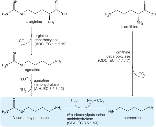
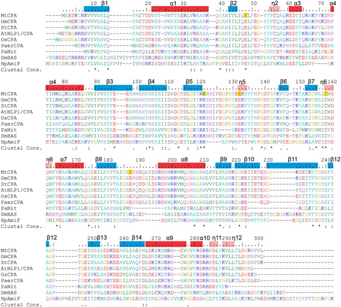
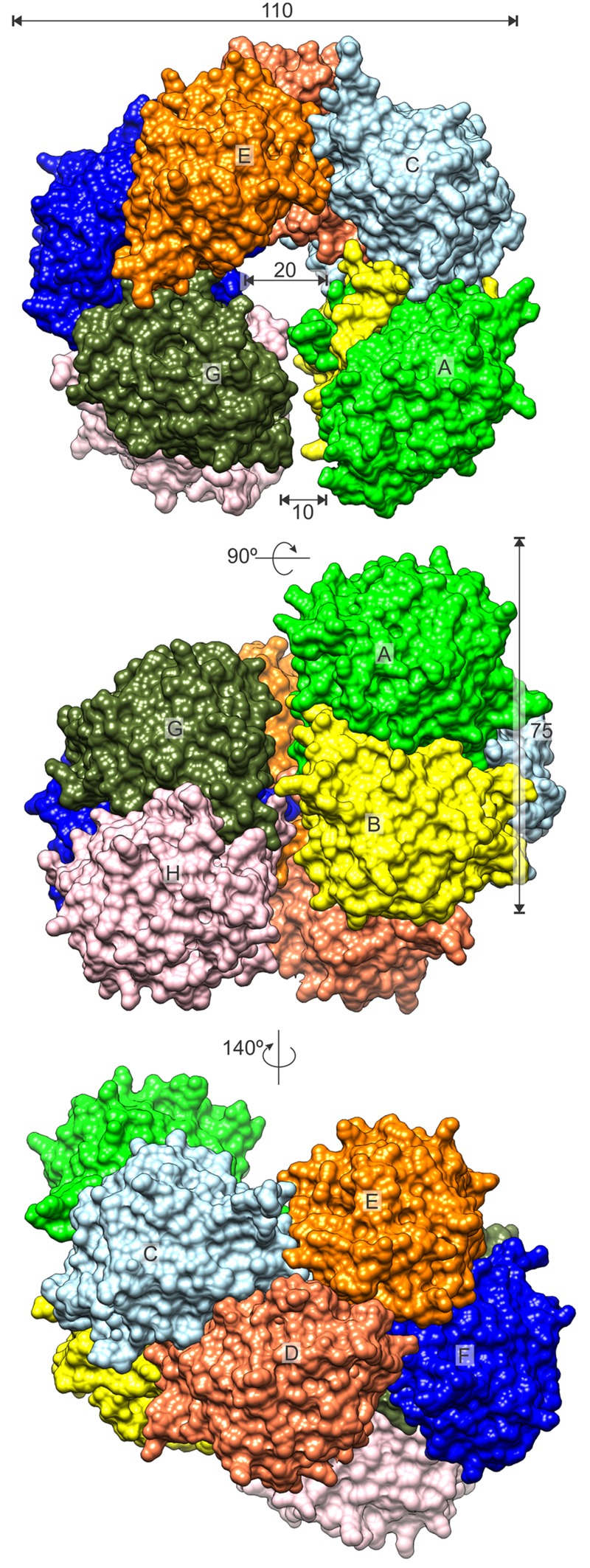
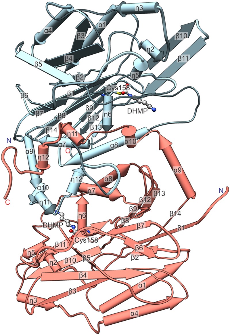
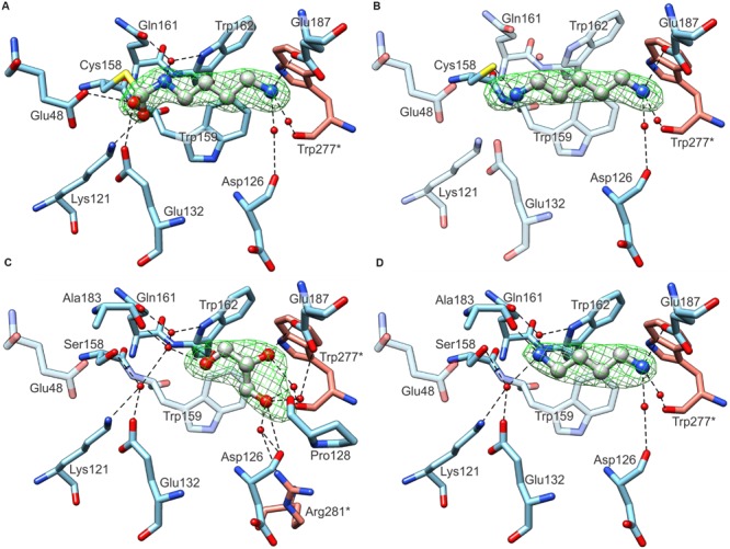
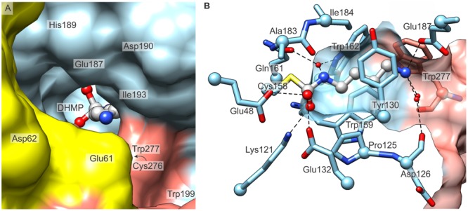
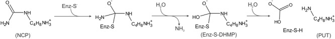
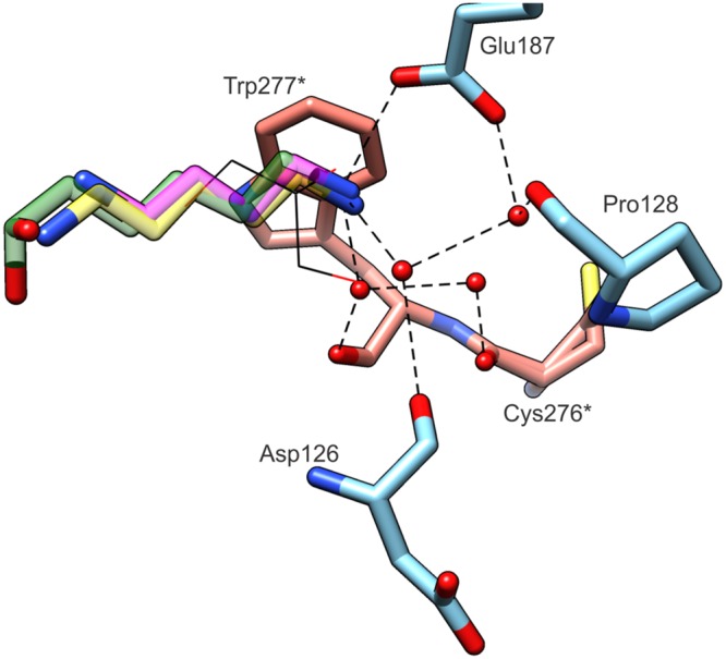
Putrescine, 1,4-diaminobutane, is an intermediate in the biosynthesis of more complexed polyamines, spermidine and spermine. Unlike other eukaryotes, plants have evolved a multistep pathway for putrescine biosynthesis that utilizes arginine. In the final reaction, N-carbamoylputrescine is hydrolyzed to putrescine by N-carbamoylputrescine amidohydrolase (CPA, EC 3.5.1.53). During the hydrolysis, consecutive nucleophilic attacks on the substrate by Cys158 and water lead to formation of putrescine and two by-products, ammonia and carbon dioxide. CPA from the model legume plant, Medicago truncatula (MtCPA), was investigated in this work. Four crystal structures were determined: the wild-type MtCPA in complex with the reaction intermediate, N-(dihydroxymethyl)putrescine as well as with cadaverine, which is a longer analog of putrescine; and also structures of MtCPA-C158S mutant unliganded and with putrescine. MtCPA assembles into octamers, which resemble an incomplete left-handed helical twist. The active site of MtCPA is funnel-like shaped, and its entrance is walled with a contribution of the neighboring protein subunits. Deep inside the catalytic cavity, Glu48, Lys121, and Cys158 form the catalytic triad. In this studies, we have highlighted the key residues, highly conserved among the plant kingdom, responsible for the activity and selectivity of MtCPA toward N-carbamoylputrescine. Moreover, since, according to previous reports, a close MtCPA relative from Arabidopsis thaliana, along with several other nitrilase-like proteins, are subjected to allosteric regulation by substrates, we have used the structural information to indicate a putative secondary binding site. Based on the docking experiment, we postulate that this site is adjacent to the entrance to the catalytic pocket.

Keywords: 1; 4-diaminobutane; CPA; amidase mechanism; cadaverine; carbamoyl hydrolysis; crystal structure; octamer; polyamine synthesis.


Figures

FIGURE 1
FIGURE 1
Two pathways for putrescine biosynthesis. The reaction catalyzed by MtCPA is highlighted in blue.
FIGURE 2
FIGURE 2
Sequence alignment of selected nitrilase enzymes. UniProt accession numbers are given in square brackets, and the values in per cent indicate sequence identity to MtCPA. Six first sequences correspond to CPA enzymes from: MtCPA [G7ITU5]; Glycine max, (GmCPA) [I1M4B9] 94%; Solanum tuberosum, (StCPA) [Q3HVN1] 86%; Arabidopsis thaliana, (AtNLP1/CPA) [B9DGV9] 85%; Os Indica group putative protein (OsCPA) [A2X5P5] 82%, Pseudomonas aeruginosa CPA (PaerCPA) [A6UY94] 64%. The other three sequences belong to protein structures described in this manuscript: Pyrococcus abyssi Nitrilase (PaNit) [Q9UYV8] 35%; Drosophila melanogaster β-Alanine Synthase without 60 N-terminal residues (DmβAS) [Q9VI04] 31% and Helicobacter pylori Formamidase (HpAmiF) [M3MZ63] 27%. Numbering above the sequences and annotation of the secondary structure elements (α helices, red bars; η 310 helices, pink; β strands, blue) corresponds to MtCPA. Residues are color-coded by type, and E48, K121, E132, C158 and E187 in MtCPA sequence are highlighted in yellow.
FIGURE 3
FIGURE 3
MtCPA octamer. Surface of each protein subunit (capital letters, A–H) is colored differently. Dimensions are given in Å.
FIGURE 4
FIGURE 4
Detailed view on an isolated MtCPA dimer with annotation of the secondary structure. Numbering of helices is consecutive, regardless of their type, and η indicate 310 helices. The reaction intermediate, DHMP and the active Cys158 are pictured to visualize the enzymatic reaction venue. Protein chains C (light blue) and D (salmon) are shown.
FIGURE 5
FIGURE 5
Ligand binding by MtCPA (A,B) and MtCPA C158S mutant (C,D). In each panel protein subunits C (blue C atoms) and D (salmon) are shown whereas the ligands: DHMP in (A); CAD (B); PUT (C) and GOL (D) are in gray. Green meshes visualize omit electron density maps contoured at 5 σ level around ligands. Dashed black lines indicate hydrogen bonds. Semitransparent residues in (B–D) do not interact with ligands, and are depicted for comparison only.
FIGURE 6
FIGURE 6
The active site of MtCPA. (A) View along the entrance to the cavity which contains the active site, and the reaction intermediate, DHMP inside. Protein subunits B (yellow), C (light blue) and D (salmon) are shown. (B) Detailed mode of interaction of MtCPA with DHMP. Dashed lines indicate hydrogen-bonded atoms. Hydrogen bonds mediated by more than one water molecule are omitted for clarity. Cα atoms of interacting residues are shown as balls. The protein surface (semitransparent, C and D) is clipped to show the maximum vista over the binding cavity.
FIGURE 7
FIGURE 7
A scheme of enzymatic hydrolysis of NCP to PUT. Enz-S-DHMP indicates the covalent complex with N-(dihydroxymethyl)putrescine, observed in the MtCPA/DHMP structure.
FIGURE 8
FIGURE 8
Substrate specificity of MtCPA. The ligands (semitransparent) inside the cavity originate from MtCPA/DHMP complex (green); MtCPA/CAD (yellow); MtCPA-C158S/PUT (magenta). GOL molecule (black wires) is superposed from MtCPA-C158S unliganded structure. Interacting amino acid residues and hydrogen-bonding network (dashed black lines) are shown for MtCPA/DHMP complex only, but their locations are preserved in MtCPA/CAD and MtCPA-C158S/PUT complexes as well. Asterisks () indicate amino acid residues that belong to dimer-mate subunit. Interactions mediated by more than two water molecules (red balls) are omitted for clarity.
FIGURE 9
FIGURE 9
The proposed alternative substrate-binding site. The substrate (ball-and-sticks, green C atoms) was docked at the location where a GOL molecule (magenta) is present universally in all presented crystal structures. Note that the carbamoyl moiety of the substrate partially overlaps with the GOL position. The reaction intermediate, DHMP (ball-and-sticks, gray), bound in the catalytic center is shown to visualize the structural proximity of the two sites. Dashed black lines indicate possible hydrogen bonds with Asp194, His198 and Glu248. The surfaces of the two dimer-forming protein subunits are color-coded red and blue.

References

    1. Agarkar V. B., Kimani S. W., Cowan D. A., Sayed M. F., Sewell B. T. (2006). The quaternary structure of the amidase from Geobacillus pallidus RAPc8 is revealed by its crystal packing. Acta Crystallogr. Sect. F Struct. Biol. Cryst. Commun. 62 1174–1178. 10.1107/S1744309106043855 - DOI - PMC - PubMed
    1. Alcazar R., Planas J., Saxena T., Zarza X., Bortolotti C., Cuevas J., et al. (2010). Putrescine accumulation confers drought tolerance in transgenic Arabidopsis plants over-expressing the homologous Arginine decarboxylase 2 gene. Plant Physiol. Biochem. 48 547–552. 10.1016/j.plaphy.2010.02.002 - DOI - PubMed
    1. Bachrach U. (2010). The early history of polyamine research. Plant Physiol. Biochem. 48 490–495. 10.1016/j.plaphy.2010.02.003 - DOI - PubMed
    1. Barglow K. T., Saikatendu K. S., Bracey M. H., Huey R., Morris G. M., Olson A. J., et al. (2008). Functional proteomic and structural insights into molecular recognition in the nitrilase family enzymes. Biochemistry 47 13514–13523. 10.1021/bi801786y - DOI - PMC - PubMed
    1. Berman H. M., Westbrook J., Feng Z., Gilliland G., Bhat T. N., Weissig H., et al. (2000). The protein data bank. Nucleic Acids Res. 28 235–242. 10.1093/nar/28.1.235 - DOI - PMC - PubMed

LinkOut - more resources