

Expression of perilipin 5 promotes lipid droplet formation in yeast
- PMID: 27066172
- PMCID: PMC4802777
- DOI: 10.1080/19420889.2015.1071728
Expression of perilipin 5 promotes lipid droplet formation in yeast
Abstract
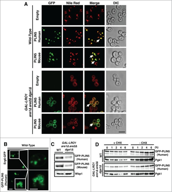
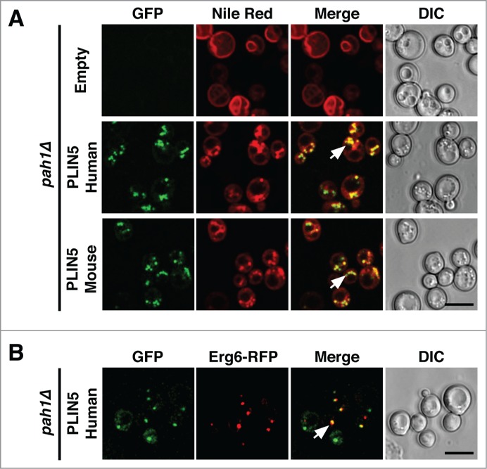
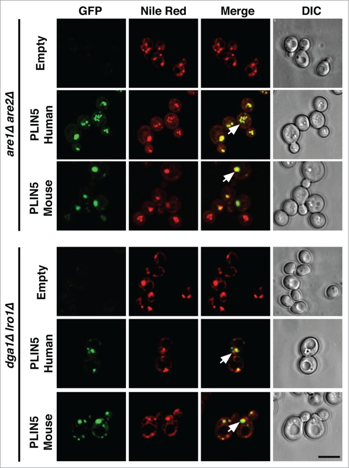
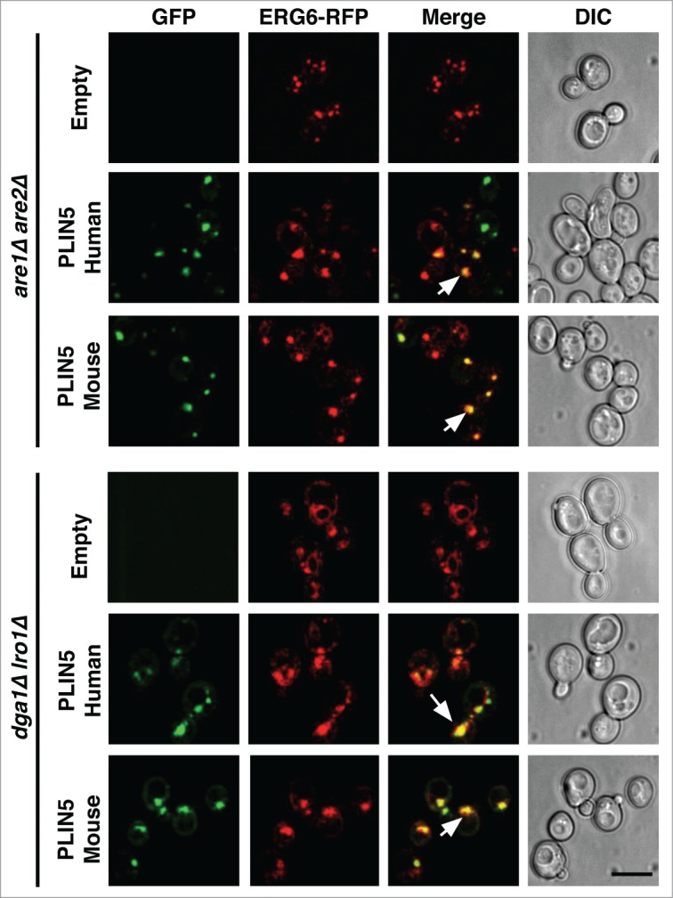
Neutral lipids are packed into dedicated intracellular compartments termed lipid droplets (LDs). LDs are spherical structures delineated by an unusual lipid monolayer and they harbor a specific set of proteins, many of which function in lipid synthesis and lipid turnover. In mammals, LDs are covered by abundant scaffolding proteins, the perilipins (PLIN1-5). LDs in yeast are functionally similar to that of mammalian cells, but they lack the perilipins. We have previously shown that perilipins (PLIN1-3) are properly targeted to LDs when expressed in yeast and that they promote LD formation from the ER membrane enriched in neutral lipids. Here we address the question whether PLIN5 (OXPAT) has a similar function. Both human and murine PLIN5 were properly targeted to yeast LDs, but the protein localized to the cytosol and its steady-state level was reduced when expressed in yeast mutants lacking the capacity to synthesize storage lipids. When expressed in cells containing high levels of neutral lipids within the membrane of the endoplasmatic reticulum, PLIN5 promoted the formation of LDs. Interestingly, PLIN5 was properly targeted to LDs, irrespective of whether these LDs were filled with triacylglycerol or steryl esters, indicating that PLIN5 did not exhibit targeting specificity for a particular subtypes of LDs as was reported for mammalian cells.
Keywords: PLIN5 (OXPAT); Saccharomyces cerevisiae; endoplasmatic reticulum; lipid droplets; organelle biogenesis; perilipins; steryl esters; triacylglycerol.
Figures
Similar articles
-
Expression of oleosin and perilipins in yeast promotes formation of lipid droplets from the endoplasmic reticulum.J Cell Sci. 2013 Nov 15;126(Pt 22):5198-209. doi: 10.1242/jcs.131896. Epub 2013 Sep 4. J Cell Sci. 2013. PMID: 24006263
-
Perilipin 3 promotes the formation of membrane domains enriched in diacylglycerol and lipid droplet biogenesis proteins.Front Cell Dev Biol. 2023 Jul 3;11:1116491. doi: 10.3389/fcell.2023.1116491. eCollection 2023. Front Cell Dev Biol. 2023. PMID: 37465010 Free PMC article.
-
The surface of lipid droplets constitutes a barrier for endoplasmic reticulum-resident integral membrane proteins.J Cell Sci. 2022 Mar 1;135(5):jcs256206. doi: 10.1242/jcs.256206. Epub 2021 May 24. J Cell Sci. 2022. PMID: 34028531
-
Molecular mechanisms of perilipin protein function in lipid droplet metabolism.FEBS Lett. 2024 May;598(10):1170-1198. doi: 10.1002/1873-3468.14792. Epub 2024 Jan 1. FEBS Lett. 2024. PMID: 38140813 Review.
-
Pathophysiology of lipid droplet proteins in liver diseases.Exp Cell Res. 2016 Jan 15;340(2):187-92. doi: 10.1016/j.yexcr.2015.10.021. Epub 2015 Oct 26. Exp Cell Res. 2016. PMID: 26515554 Free PMC article. Review.
Cited by
-
Mouse fat storage-inducing transmembrane protein 2 (FIT2) promotes lipid droplet accumulation in plants.Plant Biotechnol J. 2017 Jul;15(7):824-836. doi: 10.1111/pbi.12678. Epub 2017 Jan 18. Plant Biotechnol J. 2017. PMID: 27987528 Free PMC article.
-
Proteomics Analysis of Lipid Droplets from the Oleaginous Alga Chromochloris zofingiensis Reveals Novel Proteins for Lipid Metabolism.Genomics Proteomics Bioinformatics. 2019 Jun;17(3):260-272. doi: 10.1016/j.gpb.2019.01.003. Epub 2019 Sep 5. Genomics Proteomics Bioinformatics. 2019. PMID: 31494267 Free PMC article.
-
Interdigitation between Triglycerides and Lipids Modulates Surface Properties of Lipid Droplets.Biophys J. 2017 Apr 11;112(7):1417-1430. doi: 10.1016/j.bpj.2017.02.032. Biophys J. 2017. PMID: 28402884 Free PMC article.
References
-
- Walther TC, Farese RVJ. Lipid droplets and cellular lipid metabolism. Annu Rev Biochem 2012; 81:687-714; PMID:22524315; http://dx.doi.org/10.1146/annurev-biochem-061009-102430 - DOI - PMC - PubMed
-
- Jacquier N, Choudhary V, Mari M, Toulmay A, Reggiori F, Schneiter R. Lipid droplets are functionally connected to the endoplasmic reticulum in Saccharomyces cerevisiae. J Cell Sci 2011; 124:2424-37; PMID:21693588; http://dx.doi.org/10.1242/jcs.076836 - DOI - PubMed
-
- Wilfling F, Wang H, Haas JT, Krahmer N, Gould TJ, Uchida A, Cheng JX, Graham M, Christiano R, Fröhlich F, et al.. Triacylglycerol synthesis enzymes mediate lipid droplet growth by relocalizing from the ER to lipid droplets. Dev Cell 2013; 24:384-99; PMID:23415954; http://dx.doi.org/10.1016/j.devcel.2013.01.013 - DOI - PMC - PubMed
-
- Kimmel AR, Brasaemle DL, McAndrews-Hill M, Sztalryd C, Londos C. Adoption of PERILIPIN as a unifying nomenclature for the mammalian PAT-f amily of intracellular lipid storage droplet proteins. J Lipid Res 2010; 51:468-71; PMID:19638644; http://dx.doi.org/10.1194/jlr.R000034 - DOI - PMC - PubMed
-
- Brasaemle DL. Thematic review series: adipocyte biology. The perilipin family of structural lipid droplet proteins: stabilization of lipid droplets and control of lipolysis. J Lipid Res 2007; 48:2547-59; PMID:17878492; http://dx.doi.org/10.1194/jlr.R700014-JLR200 - DOI - PubMed
LinkOut - more resources
Full Text Sources
Other Literature Sources
Research Materials