

The challenge of ORF1p phosphorylation: Effects on L1 activity and its host
- PMID: 27066302
- PMCID: PMC4802804
- DOI: 10.1080/2159256X.2015.1119927
The challenge of ORF1p phosphorylation: Effects on L1 activity and its host
Abstract
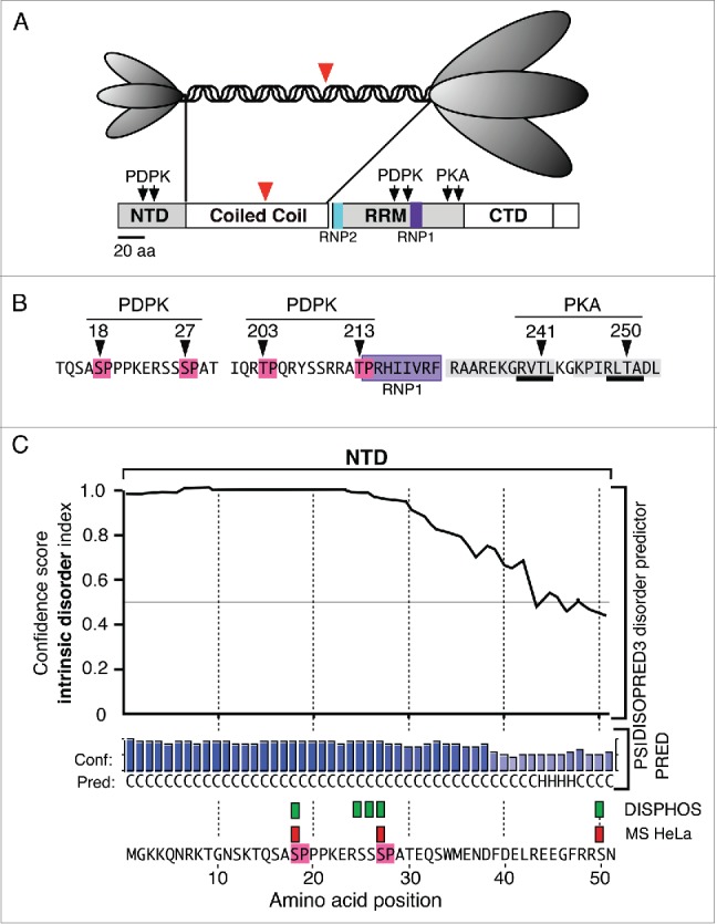
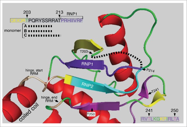
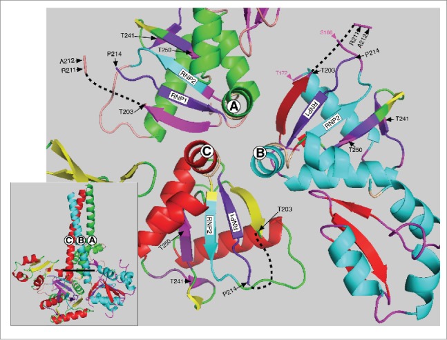
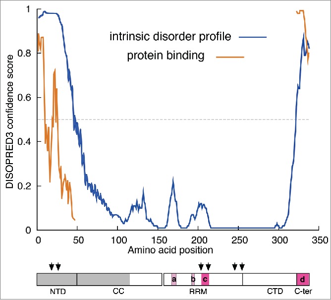
L1 non-LTR retrotransposons are autonomously replicating genetic elements that profoundly affected their mammalian hosts having generated upwards of 40% or more of their genomes. Although deleterious, they remain active in most mammalian species, and thus the nature and consequences of the interaction between L1 and its host remain major issues for mammalian biology. We recently showed that L1 activity requires phosphorylation of one of its 2 encoded proteins, ORF1p, a nucleic acid chaperone and the major component of the L1RNP retrotransposition intermediate. Reversible protein phosphorylation, which is effected by interacting cascades of protein kinases, phosphatases, and ancillary proteins, is a mainstay in the regulation and coordination of many basic biological processes. Therefore, demonstrating phosphorylation-dependence of L1 activity substantially enlarged our knowledge of the scope of L1 / host interaction. However, developing a mechanistic understanding of what this means for L1 or its host is a formidable challenge, which we discuss here.
Keywords: L1 retrotransposon; Pin1; intrinsically disordered region; peptidyl prolyl isomerase 1; proline-directed protein kinase.
Figures
Similar articles
-
Phosphorylation of ORF1p is required for L1 retrotransposition.Proc Natl Acad Sci U S A. 2015 Apr 7;112(14):4298-303. doi: 10.1073/pnas.1416869112. Epub 2015 Mar 23. Proc Natl Acad Sci U S A. 2015. PMID: 25831499 Free PMC article.
-
Nucleic acid chaperone properties of ORF1p from the non-LTR retrotransposon, LINE-1.RNA Biol. 2010 Nov-Dec;7(6):706-11. doi: 10.4161/rna.7.6.13766. Epub 2010 Nov 1. RNA Biol. 2010. PMID: 21045547 Free PMC article. Review.
-
Detection of the LINE-1 retrotransposon RNA-binding protein ORF1p in different anatomical regions of the human brain.Mob DNA. 2017 Nov 22;8:17. doi: 10.1186/s13100-017-0101-4. eCollection 2017. Mob DNA. 2017. PMID: 29201157 Free PMC article.
-
LINE-1 retrotransposition requires the nucleic acid chaperone activity of the ORF1 protein.J Mol Biol. 2005 May 6;348(3):549-61. doi: 10.1016/j.jmb.2005.03.003. J Mol Biol. 2005. PMID: 15826653
-
Protein-nucleic acid interactions of LINE-1 ORF1p.Semin Cell Dev Biol. 2019 Feb;86:140-149. doi: 10.1016/j.semcdb.2018.03.019. Epub 2018 Mar 31. Semin Cell Dev Biol. 2019. PMID: 29596909 Free PMC article. Review.
Cited by
-
LINE-1 ribonucleoprotein condensates bind DNA to enable nuclear entry during mitosis.Sci Adv. 2025 May 2;11(18):eadt9318. doi: 10.1126/sciadv.adt9318. Epub 2025 May 2. Sci Adv. 2025. PMID: 40315332 Free PMC article.
-
Diverse single-stranded nucleic acid binding proteins enable both stable protection and rapid exchange required for biological function.QRB Discov. 2025 Jan 14;6:e1. doi: 10.1017/qrd.2024.21. eCollection 2025. QRB Discov. 2025. PMID: 39944884 Free PMC article.
-
L1-ORF1p nucleoprotein can rapidly assume distinct conformations and simultaneously bind more than one nucleic acid.Nucleic Acids Res. 2024 Dec 11;52(22):14013-14029. doi: 10.1093/nar/gkae1141. Nucleic Acids Res. 2024. PMID: 39565204 Free PMC article.
-
Melatonin: Regulation of Viral Phase Separation and Epitranscriptomics in Post-Acute Sequelae of COVID-19.Int J Mol Sci. 2022 Jul 23;23(15):8122. doi: 10.3390/ijms23158122. Int J Mol Sci. 2022. PMID: 35897696 Free PMC article. Review.
-
Co-expression of distinct L1 retrotransposon coiled coils can lead to their entanglement.Mob DNA. 2023 Oct 20;14(1):16. doi: 10.1186/s13100-023-00303-8. Mob DNA. 2023. PMID: 37864180 Free PMC article.
References
-
- Moran JV, Holmes SE, Naas TP, DeBerardinis RJ, Boeke JD, Kazazian HH, Jr. High frequency retrotransposition in cultured mammalian cells. Cell 1996; 7(5):917-27; PMID:8945518; http://dx.doi.org/10.1016/S0092-8674(00)81998-4 - DOI - PubMed
-
- Martin SL. Ribonucleoprotein particles with LINE-1 RNA in mouse embryonal carcinoma cells. Mol Cell Biol 1991; 11(9):4804-7; PMID:1715025; http://dx.doi.org/10.1128/MCB.11.9.4804 - DOI - PMC - PubMed
-
- Wei W, Gilbert N, Ooi SL, Lawler JF, Ostertag EM, Kazazian HH, Boeke JD, Moran JV. Human L1 retrotransposition: cis preference versus trans complementation. Mol Cell Biol 2001; 21(4):1429-39; PMID:11158327; http://dx.doi.org/10.1128/MCB.21.4.1429-1439.2001 - DOI - PMC - PubMed
-
- Kulpa DA, Moran JV. Ribonucleoprotein particle formation is necessary but not sufficient for LINE-1 retrotransposition. Hum Mol Genet 2005; 14(21):3237-48; PMID:16183655; http://dx.doi.org/10.1093/hmg/ddi354 - DOI - PubMed
LinkOut - more resources
Full Text Sources
Miscellaneous