

Cachexia-associated adipose tissue morphological rearrangement in gastrointestinal cancer patients
- PMID: 27066317
- PMCID: PMC4799865
- DOI: 10.1002/jcsm.12037
Cachexia-associated adipose tissue morphological rearrangement in gastrointestinal cancer patients
Abstract
Background and aims: Cachexia is a syndrome characterized by marked involuntary loss of body weight. Recently, adipose tissue (AT) wasting has been shown to occur before the appearance of other classical cachexia markers. We investigated the composition and rearrangement of the extracellular matrix, adipocyte morphology and inflammation in the subcutaneous AT (scAT) pad of gastrointestinal cancer patients.
Methods: Surgical biopsies for scAT were obtained from gastrointestinal cancer patients, who were signed up into the following groups: cancer cachexia (CC, n = 11), weight-stable cancer (WSC, n = 9) and weight-stable control (non-cancer) (control, n = 7). The stable weight groups were considered as those with no important weight change during the last year and body mass index <25 kg/m(2). Subcutaneous AT fibrosis was quantified and characterized by quantitative PCR, histological analysis and immunohistochemistry.
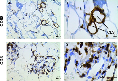
Results: The degree of fibrosis and the distribution and collagen types (I and III) were different in WSC and CC patients. CC patients showed more pronounced fibrosis in comparison with WSC. Infiltrating macrophages surrounding adipocytes and CD3 Ly were found in the fibrotic areas of scAT. Subcutaneous AT fibrotic areas demonstrated increased monocyte chemotactic protein 1 (MCP-1) and Cluster of Differentiation (CD)68 gene expression in cancer patients.
Conclusions: Our data indicate architectural modification consisting of fibrosis and inflammatory cell infiltration in scAT as induced by cachexia in gastrointestinal cancer patients. The latter was characterized by the presence of macrophages and lymphocytes, more evident in the fibrotic areas. In addition, increased MCP-1 and CD68 gene expression in scAT from cancer patients may indicate an important role of these markers in the early phases of cancer.
Keywords: Adipose tissue; Extracellular matrix; Fibrosis, Inflammation, Cachexia.
Figures


References
-
- Argiles JM, Anker SD, Evans WJ, Morley JE, Fearon KC, Strasser F, et al. Consensus on cachexia definitions. J Am Med Dir Assoc 2010; 11: 229–230. - PubMed
-
- Fearon K, Strasser F, Anker SD, Bosaeus I, Bruera E, Fainsinger RL, et al Definition and classification of cancer cachexia: an international consensus. Lancet Oncol 2011; 12: 489–495. - PubMed
-
- Tisdale MJ. Cachexia in cancer patients. Nat Rev Cancer 2002; 2: 862–871. - PubMed
-
- Fearon K, Arends J, Baracos V. Understanding the mechanisms and treatment options in cancer cachexia. Nat Rev Clin Oncol 2013; 10: 90–99. - PubMed
-
- Dodson S, Baracos VE, Jatoi A, Evans WJ, Cella D, Dalton JT, et al. Muscle wasting in cancer cachexia: clinical implications, diagnosis, and emerging treatment strategies. Annu Rev Med 2011; 62: 265–279. - PubMed
LinkOut - more resources
Full Text Sources
Other Literature Sources
Research Materials
Miscellaneous
