

Application of Arterial Spin Labelling in the Assessment of Ocular Tissues
- PMID: 27066501
- PMCID: PMC4811053
- DOI: 10.1155/2016/6240504
Application of Arterial Spin Labelling in the Assessment of Ocular Tissues
Abstract
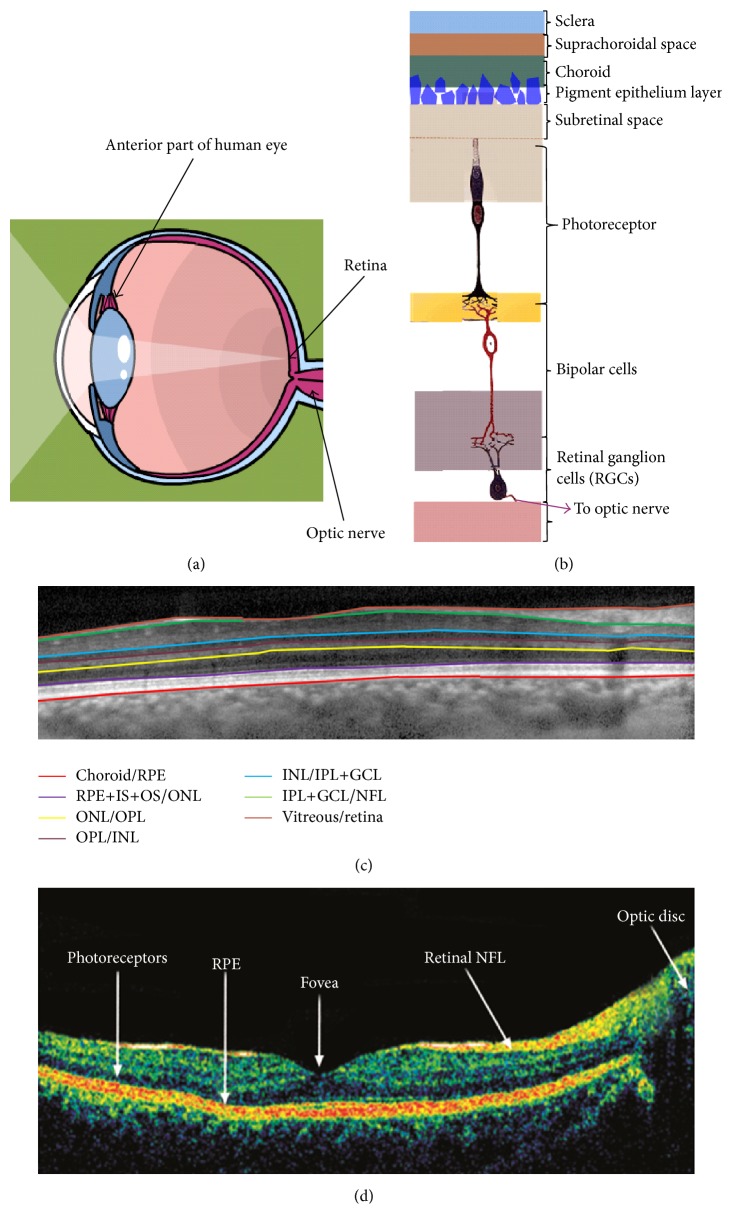
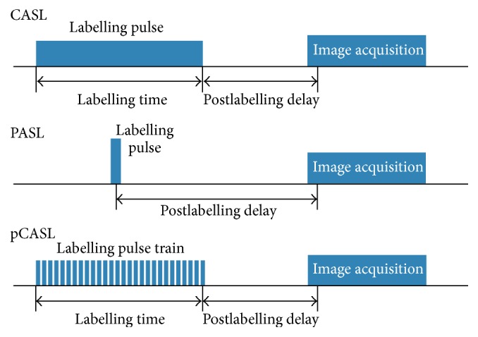
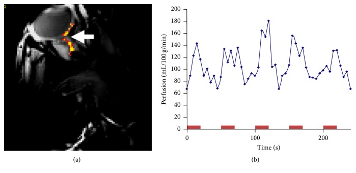
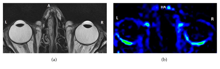
Arterial spin labelling (ASL) is a noninvasive magnetic resonance imaging (MRI) modality, capable of measuring blood perfusion without the use of a contrast agent. While ASL implementation for imaging the brain and monitoring cerebral blood flow has been reviewed in depth, the technique is yet to be widely used for ocular tissue imaging. The human retina is a very thin but highly stratified structure and it is also situated close to the surface of the body which is not ideal for MR imaging. Hence, the application of MR imaging and ASL in particular has been very challenging for ocular tissues and retina. That is despite the fact that almost all of retinal pathologies are accompanied by blood perfusion irregularities. In this review article, we have focused on the technical aspects of the ASL and their implications for its optimum adaptation for retinal blood perfusion monitoring. Retinal blood perfusion has been assessed through qualitative or invasive quantitative methods but the prospect of imaging flow using ASL would increase monitoring and assessment of retinal pathologies. The review provides details of ASL application in human ocular blood flow assessment.
Figures
Similar articles
-
Repeatability of Arterial Spin Labeling MRI in Measuring Blood Perfusion in the Human Eye.J Magn Reson Imaging. 2019 Apr;49(4):966-974. doi: 10.1002/jmri.26323. Epub 2018 Sep 25. J Magn Reson Imaging. 2019. PMID: 30252997
-
MR angiography and arterial spin labelling.Methods Mol Biol. 2011;711:327-45. doi: 10.1007/978-1-61737-992-5_16. Methods Mol Biol. 2011. PMID: 21279610
-
Arterial spin-labeling MR imaging in Moyamoya disease compared with clinical assessments and other MR imaging findings.Eur J Radiol. 2013 Dec;82(12):e840-7. doi: 10.1016/j.ejrad.2013.08.040. Epub 2013 Sep 1. Eur J Radiol. 2013. PMID: 24055185
-
Arterial spin labeling in clinical pediatric imaging.Diagn Interv Imaging. 2016 Feb;97(2):151-8. doi: 10.1016/j.diii.2015.09.001. Epub 2015 Oct 9. Diagn Interv Imaging. 2016. PMID: 26456912 Review.
-
Mapping of cerebral perfusion territories using territorial arterial spin labeling: techniques and clinical application.NMR Biomed. 2013 Aug;26(8):901-12. doi: 10.1002/nbm.2836. Epub 2012 Jul 15. NMR Biomed. 2013. PMID: 22807022 Review.
Cited by
-
ABCB1 and ABCG2 Together Limit the Distribution of ABCB1/ABCG2 Substrates to the Human Retina and the ABCG2 Single Nucleotide Polymorphism Q141K (c.421C> A) May Lead to Increased Drug Exposure.Front Pharmacol. 2021 Jun 16;12:698966. doi: 10.3389/fphar.2021.698966. eCollection 2021. Front Pharmacol. 2021. PMID: 34220523 Free PMC article.
-
Application of Arterial Spin Labelling in Detecting Retinal Ischemia.Case Rep Ophthalmol. 2017 Dec 14;8(3):545-557. doi: 10.1159/000485316. eCollection 2017 Sep-Dec. Case Rep Ophthalmol. 2017. PMID: 29422857 Free PMC article.
-
In Vivo Detection of the Effect of Electroacupuncture on "Zusanli" Acupoint in Rats with Adjuvant-Induced Arthritis through Optical Coherence Tomography.Biomed Res Int. 2016;2016:2681463. doi: 10.1155/2016/2681463. Epub 2016 Nov 17. Biomed Res Int. 2016. PMID: 27981046 Free PMC article.
References
-
- Reynolds J. D., Olitsky S. E. Pediatric Retina. Springer; 2010.
-
- Lens A., Nemeth S. C., Ledford J. K. Ocular Anatomy and Physiology. Thorofare, NJ, USA: SLACK Incorporated; 2008.
-
- Abramson D. Blood Vessels and Lymphatics in Organ Systems. Academic Press; 2012.
-
- Zhang H. R. Scanning electron-microscopic study of corrosion casts on retinal and choroidal angioarchitecture in man and animals. Progress in Retinal and Eye Research. 1994;13(1):243–270. doi: 10.1016/1350-9462(94)90012-4. - DOI
-
- Kiel J. W. The Ocular Circulation. San Rafael, Calif, USA: Morgan & Claypool Life Sciences; 2010. (Integrated Systems Physiology: From Molecule to Function to Disease). - PubMed
Publication types
MeSH terms
Substances
LinkOut - more resources
Full Text Sources
Other Literature Sources
Medical
