

The Neuropsychiatric Disease-Associated Gene cacna1c Mediates Survival of Young Hippocampal Neurons
- PMID: 27066530
- PMCID: PMC4819284
- DOI: 10.1523/ENEURO.0006-16.2016
The Neuropsychiatric Disease-Associated Gene cacna1c Mediates Survival of Young Hippocampal Neurons
Abstract
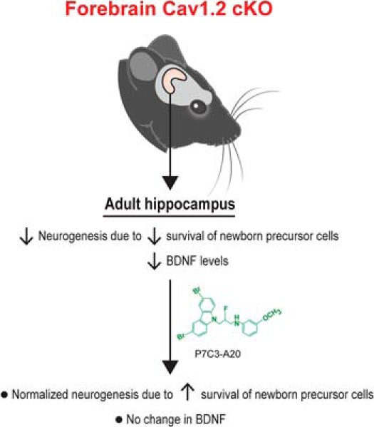
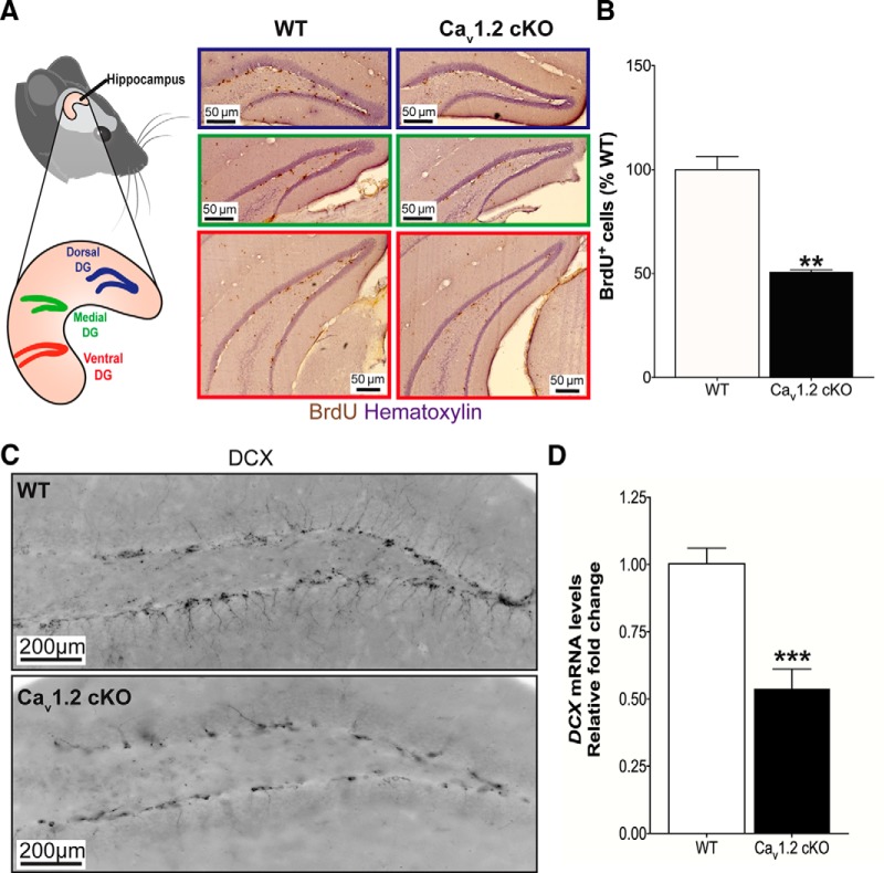
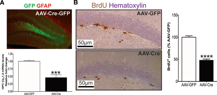
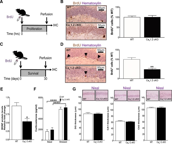
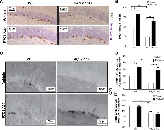
Genetic variations in CACNA1C, which encodes the Cav1.2 subunit of L-type calcium channels (LTCCs), are associated with multiple forms of neuropsychiatric disease that manifest high anxiety in patients. In parallel, mice harboring forebrain-specific conditional knockout of cacna1c (forebrain-Cav1.2 cKO) display unusually high anxiety-like behavior. LTCCs in general, including the Cav1.3 subunit, have been shown to mediate differentiation of neural precursor cells (NPCs). However, it has not previously been determined whether Cav1.2 affects postnatal hippocampal neurogenesis in vivo. Here, we show that forebrain-Cav1.2 cKO mice exhibit enhanced cell death of young hippocampal neurons, with no change in NPC proliferation, hippocampal size, dentate gyrus thickness, or corticosterone levels compared with wild-type littermates. These mice also exhibit deficits in brain levels of brain-derived neurotrophic factor (BDNF), and Cre recombinase-mediated knockdown of adult hippocampal Cav1.2 recapitulates the deficit in young hippocampal neurons survival. Treatment of forebrain-Cav1.2 cKO mice with the neuroprotective agent P7C3-A20 restored the net magnitude of postnatal hippocampal neurogenesis to wild-type levels without ameliorating their deficit in BDNF expression. The role of Cav1.2 in young hippocampal neurons survival may provide new approaches for understanding and treating neuropsychiatric disease associated with aberrations in CACNA1C. Visual Abstract.
Keywords: Cav; P7C3; P7C3A20; anxiety; neurogenesis; neuroprotection.
Figures
Similar articles
-
Cacna1c: Protecting young hippocampal neurons in the adult brain.Neurogenesis (Austin). 2016 Sep 2;3(1):e1231160. doi: 10.1080/23262133.2016.1231160. eCollection 2016. Neurogenesis (Austin). 2016. PMID: 27900342 Free PMC article.
-
The L-type calcium channel Cav1.3 is required for proper hippocampal neurogenesis and cognitive functions.Cell Calcium. 2015 Dec;58(6):606-16. doi: 10.1016/j.ceca.2015.09.007. Epub 2015 Oct 3. Cell Calcium. 2015. PMID: 26459417
-
Deletion of the Mouse Homolog of CACNA1C Disrupts Discrete Forms of Hippocampal-Dependent Memory and Neurogenesis within the Dentate Gyrus.eNeuro. 2016 Nov 28;3(6):ENEURO.0118-16.2016. doi: 10.1523/ENEURO.0118-16.2016. eCollection 2016 Nov-Dec. eNeuro. 2016. PMID: 27957527 Free PMC article.
-
CACNA1C: Association With Psychiatric Disorders, Behavior, and Neurogenesis.Schizophr Bull. 2018 Aug 20;44(5):958-965. doi: 10.1093/schbul/sby096. Schizophr Bull. 2018. PMID: 29982775 Free PMC article. Review.
-
L-type calcium channels and neuropsychiatric diseases: Insights into genetic risk variant-associated genomic regulation and impact on brain development.Channels (Austin). 2023 Dec;17(1):2176984. doi: 10.1080/19336950.2023.2176984. Channels (Austin). 2023. PMID: 36803254 Free PMC article. Review.
Cited by
-
Cacna1c deficiency in forebrain glutamatergic neurons alters behavior and hippocampal plasticity in female mice.Transl Psychiatry. 2024 Oct 6;14(1):421. doi: 10.1038/s41398-024-03140-2. Transl Psychiatry. 2024. PMID: 39370418 Free PMC article.
-
Downregulation of neurodevelopmental gene expression in iPSC-derived cerebral organoids upon infection by human cytomegalovirus.iScience. 2022 Mar 17;25(4):104098. doi: 10.1016/j.isci.2022.104098. eCollection 2022 Apr 15. iScience. 2022. PMID: 35391828 Free PMC article.
-
Dual-process brain mitochondria isolation preserves function and clarifies protein composition.Proc Natl Acad Sci U S A. 2021 Mar 16;118(11):e2019046118. doi: 10.1073/pnas.2019046118. Proc Natl Acad Sci U S A. 2021. PMID: 33836587 Free PMC article.
-
BDNF Levels According to Variations in the CACNA1C Gene: Sex-Based Disparity.Cell Mol Neurobiol. 2023 Jan;43(1):357-366. doi: 10.1007/s10571-022-01189-5. Epub 2022 Feb 6. Cell Mol Neurobiol. 2023. PMID: 35128618 Free PMC article.
-
Neuronal PAS Domain Proteins 1 and 3 Are Master Regulators of Neuropsychiatric Risk Genes.Biol Psychiatry. 2017 Aug 1;82(3):213-223. doi: 10.1016/j.biopsych.2017.03.021. Epub 2017 Apr 6. Biol Psychiatry. 2017. PMID: 28499489 Free PMC article.
References
-
- Ament SA, Szelinger S, Glusman G, Ashworth J, Hou L, Akula N, Shekhtman T, Badner JA, Brunkow ME, Mauldin DE, Stittrich AB, Rouleau K, Detera-Wadleigh SD, Nurnberger JI Jr, Edenberg HJ, Gershon ES, Schork N; Bipolar Genome Study. (2015) Rare variants in neuronal excitability genes influence risk for bipolar disorder. Proc Natl Acad Sci U S A 112: 3576-3581. 10.1073/pnas.1424958112 - DOI - PMC - PubMed
-
- Cameron HA, Gould E (1994) Adult neurogenesis is regulated by adrenal steroids in the dentate gyrus. Neuroscience 61: 203–209. - PubMed
Publication types
MeSH terms
Substances
Grants and funding
LinkOut - more resources
Full Text Sources
Other Literature Sources
Molecular Biology Databases