. 2015 Apr 17;1(1):e4.
doi: 10.1212/01.NXG.0000464295.65736.da. eCollection 2015 Jun.

Epileptic encephalopathy-causing mutations in DNM1 impair synaptic vesicle endocytosis

Affiliations

Epileptic encephalopathy-causing mutations in DNM1 impair synaptic vesicle endocytosis

Ryan S Dhindsa et al. Neurol Genet. .

Abstract

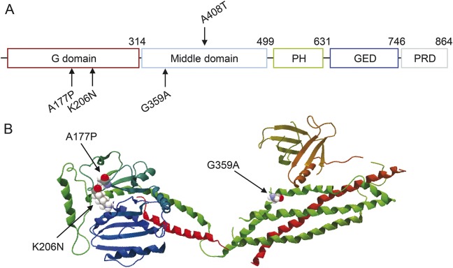
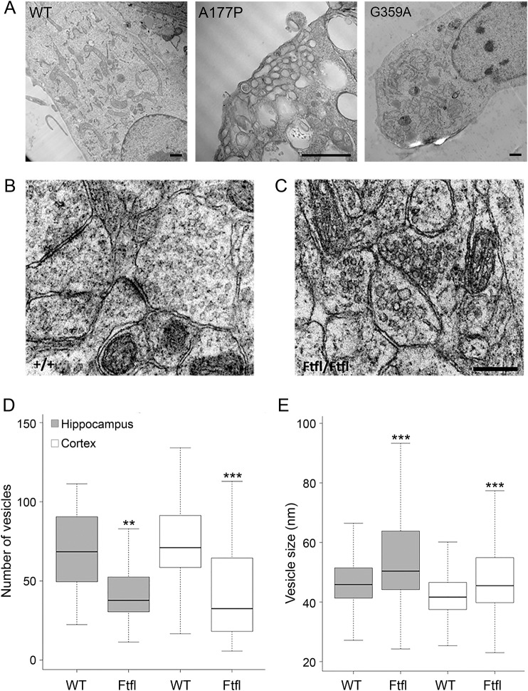
Objective: To elucidate the functional consequences of epileptic encephalopathy-causing de novo mutations in DNM1 (A177P, K206N, G359A), which encodes a large mechanochemical GTPase essential for neuronal synaptic vesicle endocytosis.

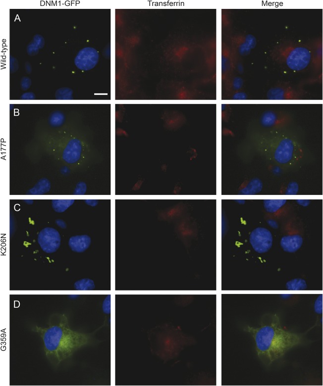
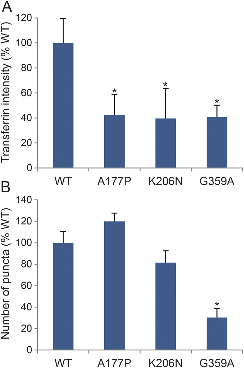
Methods: HeLa and COS-7 cells transfected with wild-type and mutant DNM1 constructs were used for transferrin assays, high-content imaging, colocalization studies, Western blotting, and electron microscopy (EM). EM was also conducted on the brain sections of mice harboring a middle-domain Dnm1 mutation (Dnm1 (Ftfl)).

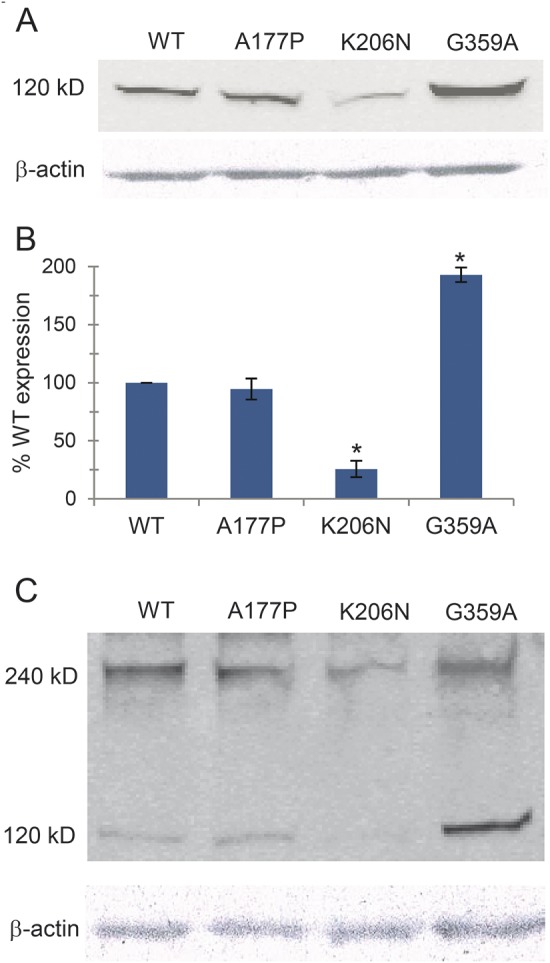
Results: We demonstrate that the expression of each mutant protein decreased endocytosis activity in a dominant-negative manner. One of the G-domain mutations, K206N, decreased protein levels. The G359A mutation, which occurs in the middle domain, disrupted higher-order DNM1 oligomerization. EM of mutant DNM1-transfected HeLa cells and of the Dnm1 (Ftfl) mouse brain revealed vesicle defects, indicating that the mutations likely interfere with DNM1's vesicle scission activity.

Conclusion: Together, these data suggest that the dysfunction of vesicle scission during synaptic vesicle endocytosis can lead to serious early-onset epilepsies.


Figures

Figure 1
Figure 1. DNM1 mutations and structure
(A) Structure-based domain architecture of human DNM1. The A177P and K206N mutations occur in the G domain and the G359A mutation occurs in the middle domain. The location of the mouse Dnm1Ftfl (A408T) mutation is indicated above the middle domain. (B) Positions of the amino acid substitutions in the DNM1 crystal structure, shown as a ribbon-type representation.
Figure 2
Figure 2. DNM1 mutations inhibit transferrin uptake
Inhibition of transferrin internalization in mammalian cell lines. COS-7 cells were transfected with green fluorescent protein (GFP)-tagged DNM1 constructs and then treated with fluorescently labeled transferrin. Scale bar, 20 μm. (A) Cells expressing wild type (WT) DNM1 exhibit transferrin uptake with a perinuclear accumulation. WT DNM1 forms round puncta that are evenly dispersed throughout the perimeter of the cell. (B) The A177P mutant inhibits transferrin uptake. DNM1 shows some diffuse GFP signal throughout the cytosol accompanied by puncta. (C) The K206N mutant also inhibits transferrin uptake and shows abnormal aggregation of DNM1. (D) The G359A mutant shows some transferrin uptake in certain cells. There is a distinct lack of puncta, and DNM1 shows a reticular GFP signal throughout the cytosol.
Figure 3
Figure 3. Quantification of transferrin uptake and cellular localization patterns
(A) High-content imaging analysis of transferrin fluorophore intensity in positively transfected HeLa cells. All 3 mutations confer nearly a 60% reduction in transferrin uptake compared to wild type (WT). Error bars represent SD between 5 replicate wells. p < 0.05 by Student t test. (B) High-content imaging spot identification revealed a 50% reduction in the number of puncta in cells expressing the G359A mutation. Error bars represent SD among 5 replicate wells. *p < 0.05 by Student t test.
Figure 4
Figure 4. DNM1 mutations affect protein levels and self-dimerization
(A) HeLa cells were transfected with green fluorescent protein (GFP)-tagged mutant constructs. The blots were probed with anti-GFP antibodies. A representative blot is shown. (B) Quantification of protein expression levels from 3 independent Western blot experiments is shown. Actin levels were used for normalization and error bars indicate SD values. *p < 0.05 by Student t test. (C) HeLa cell lysates from cells transfected with GFP-tagged mutant constructs were treated with 20 mM 1-ethyl-3-(3-dimethylaminopropyl)-carbodiimide (EDC) cross-linking agent and analyzed by Western blot. The monomeric and dimeric forms are indicated. WT = wild type.
Figure 5
Figure 5. Electron microscopy of transfected HeLa cells and Dnm1Ftfl neurons
(A) Electron microscopy (EM) of HeLa cells transfected with wild type (WT) and mutant DNM1 constructs. There are larger and abnormal vesicles in the A177P mutant. Cells transfected with the G359A show no obvious vesicle defects. Scale bars, 1 μm. (B) EM of WT mouse brain sections. (C) EM of Dnm1Ftfl mouse neurons reveals increased vesicle size. (D) A box plot depicting the number of vesicles in both WT and Dnm1Ftfl neurons. Outliers are excluded from the plot. Significance was determined by the Mann-Whitney-Wilcoxon test (**p < 0.005, ***p < 0.0005). (E) A box plot depicting the size of vesicles in both WT and Dnm1Ftfl neurons. There were significantly larger vesicles in both hippocampal and cortical neurons. Outliers are excluded from the plot. Significance was determined by the Mann-Whitney-Wilcoxon test.

References

    1. Dulac O. Epileptic encephalopathy. Epilepsia 2001;42(suppl 3):23–26. - PubMed
    1. van der Bliek AM, Redelmeier TE, Damke H, Tisdale EJ, Meyerowitz EM, Schmid SL. Mutations in human dynamin block an intermediate stage in coated vesicle formation. J Cell Biol 1993;122:553–563. - PMC - PubMed
    1. Ferguson SM, De Camilli P. Dynamin, a membrane-remodelling GTPase. Nat Rev Mol Cell Biol 2012;13:75–88. - PMC - PubMed
    1. Gray NW, Fourgeaud L, Huang B, et al. Dynamin 3 is a component of the postsynapse, where it interacts with mGluR5 and Homer. Curr Biol 2003;13:510–515. - PubMed
    1. Powell KA, Robinson PJ. Dephosphin/dynamin is a neuronal phosphoprotein concentrated in nerve terminals: evidence from rat cerebellum. Neuroscience 1995;64:821–833. - PubMed