. 2016 May 11;19(5):705-12.
doi: 10.1016/j.chom.2016.03.008. Epub 2016 Apr 5.

Type III Interferons Produced by Human Placental Trophoblasts Confer Protection against Zika Virus Infection

Affiliations

Type III Interferons Produced by Human Placental Trophoblasts Confer Protection against Zika Virus Infection

Avraham Bayer et al. Cell Host Microbe. .

Abstract

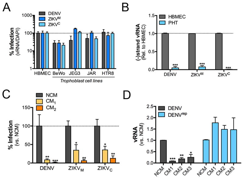
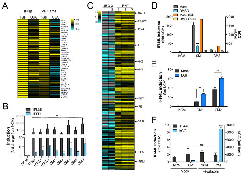
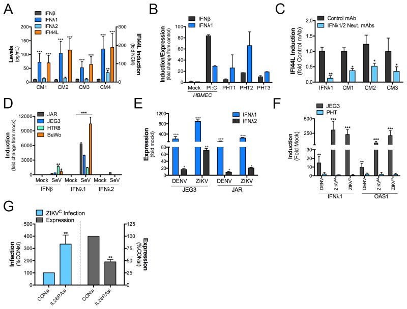
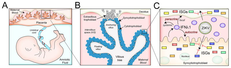
During mammalian pregnancy, the placenta acts as a barrier between the maternal and fetal compartments. The recently observed association between Zika virus (ZIKV) infection during human pregnancy and fetal microcephaly and other anomalies suggests that ZIKV may bypass the placenta to reach the fetus. This led us to investigate ZIKV infection of primary human trophoblasts (PHTs), which are the barrier cells of the placenta. We discovered that PHT cells from full-term placentas are refractory to ZIKV infection. In addition, medium from uninfected PHT cells protects non-placental cells from ZIKV infection. PHT cells constitutively release the type III interferon (IFN) IFNλ1, which functions in both a paracrine and autocrine manner to protect trophoblast and non-trophoblast cells from ZIKV infection. Our data suggest that for ZIKV to access the fetal compartment, it must evade restriction by trophoblast-derived IFNλ1 and other trophoblast-specific antiviral factors and/or use alternative strategies to cross the placental barrier.

Keywords: IFNλ; Zika virus; placenta; trophoblasts; type III interferon; virus.


Figures

Figure 1
Figure 1. ZIKV infects placental trophoblast cell lines, but not PHT cells
(A), The indicated cell lines were infected with DENV, ZIKVM, or ZIKVC for ~24hrs, fixed, and then stained with anti-dsRNA (J2) antibody. Data are shown as the percent of vRNA positive cells relative to the total number of nuclei (as assessed by DAPI). (B), Levels of DENV, ZIKVM, or ZIKVC negative strand vRNA were assessed by RT-qPCR in HBMEC or PHT cells infected for ~48hrs. (C), HBMEC were exposed to non-conditioned (NCM) PHT medium or conditioned PHT medium (CM, two independent preparations) for ~24hrs and then infected with DENV, DENV, ZIKVM, or ZIKVC. The level of infection was assessed for fluorescence microscopy for dsRNA. Data are shown as the percent of vRNA positive cells relative to the total number of nuclei (as assessed by DAPI). (D), Control HeLa cells, or HeLa cells constitutively expressing a DENV replicon were exposed to NCM or three independent preparations of PHT CM and then the levels of DENV vRNA assessed by RT-qPCR ~24hrs after exposure. In all, data are shown as mean ± standard deviation (*p<0.05, **p<0.01, ***p<0.001).
Figure 2
Figure 2. Conditioned medium from PHT cells induces ISGs
(A), A heat map of interferon stimulated genes (ISGs) differentially expressed between control (TGH) or STAT1 signaling deficient (U3A) HT1080 cells exposed to purified IFNβ or PHT CM for 24hrs. (B), RT-qPCR analysis for IFI44L or IFIT1 in U2OS cells exposed to control PHT non-conditioned medium (NCM) or five independent preparations of PHT CM. Data are shown as a fold change from NCM. (C), Heat map of differentially expressed interferon stimulated genes (ISGs) between two cultures of JEG-3 cells and a two preparations of PHT cells (samples 2 and 3 are biological replicates of the same PHT preparation) as assessed by RNASeq (p<0.05). (D), Two preparations of PHT cells were exposed to dimethyl sulfoxide (DMSO) to inhibit cell fusion, CM collected, and then IFI44L induction assessed by RT-qPCR (left y axis). In parallel, the levels of human chorionic gonadotropin (hCG) were determined by ELISA (right y axis). (E), Two preparations of PHT cells were exposed to epidermal growth factor (EGF) to enhance cell fusion, CM collected, and then IFI44L induction assessed by RT-qPCR. (F), BeWo cells were exposed to forskolin to induce fusion, CM collected, and ISG induction in CM-exposed cells assessed by RT-qPCR (for IFI44L, left y axis). In parallel, the levels of hCG were assessed by ELISA (right y axis). In (B), (D–F), data are shown as mean ± standard deviation (*p<0.05, **p<0.01, ***p<0.001, ns not significant). The color intensity in (A) and (C) indicates the level of gene expression (yellow for up-regulation and blue for down-regulation), and grey indicates that no transcripts were detected in that sample.
Figure 3
Figure 3. Conditioned medium from PHT cells contains IFNλ1, which is required for ISG induction
(A), ELISA for IFNβ, IFNλ1, and IFNλ2 in four independent PHT CM preparations (left y axis). In parallel, the extent of ISG induction in each sample was determined by RT-qPCR for the levels of IFI44L induced in U2OS cells exposed to the sample (right y axis). (B), The levels of IFNβ and IFNλ1 mRNA in three preparations of PHT cells was assessed by RT-qPCR. In parallel, IFNβ and IFNλ1 mRNA levels were determined in mock-treated HBMEC, or in HBMEC exposed to 10μg poly I:C (‘floated’ in the medium) for ~24hrs. Data are shown as a fold change from mock-treated HBMEC cells. (C), Level of ISG induction (as assessed by IFI44L RT-qPCR) in U2OS cells exposed to purified IFNλ1, or to three preparations of PHT CM incubated with a non-neutralizing monoclonal antibody (MOPC21) or anti-IFNλ-1-3 neutralizing antibodies. (D), RT-qPCR for IFNβ, IFNλ1, or IFNλ2 in indicated trophoblast cell lines infected with Sendai virus (SeV) for ~24hrs. (E), RT-qPCR for IFNλ1 or IFNλ2 in the indicated trophoblast cell lines infected with DENV or ZIKVM for ~24hrs. (F), RT-qPCR for IFNλ1 or OAS1 in JEG-3 or PHT cells infected with DENV, ZIKVM, or ZIKVC for ~24hrs. (G), ZIKVC infection in HBMEC transfected with control siRNA (CONsi) or IL28RA siRNAs and exposed to PHT conditioned medium depleted of vesicles for ~24hrs prior to infection. In all panels, data are shown as mean ± standard deviation (*p<0.05, **p<0.01, ***p<0.001, ns not significant).
Figure 4
Figure 4. Schematic depicting the structure of the human placenta and the role of IFNλ1 in protecting against ZIKV infection
(A), The intrauterine environment during human pregnancy. Embryonic structures include the villous tree of the human hemochorial placenta and the umbilical cord, which transfers blood between the placenta and the fetus. (B), An overview of a single placental villus. Extravillous trophoblasts invade and anchor the placenta to the maternal decidua and to the inner third of the myometrium. The villous tree consists of both floating and anchoring villi. Multinucleated syncytiotrophoblasts overlie the surfaces of the villous tree and are in direct contact with maternal blood, which fills the intervillous space (IVS) once the placenta is fully formed. Mononuclear cytotrophoblasts are subjacent to the syncytiotrophoblasts and the basement membrane of the villous tree, and serve to replenish the syncytiotrophoblast layer throughout pregnancy. (C), In the work presented here, we show that syncytiotrophoblasts release IFNλ1 that can act in both autocrine and paracrine manners to induce ISGs, which protect against ZIKV, and other viral infections. The paracrine function of IFNλ could work locally within the direct maternal-fetal compartment, or might circulate more systemically to act on other maternal target cells.

Comment in

References

    1. Ansarah-Sobrinho C, Nelson S, Jost CA, Whitehead SS, Pierson TC. Temperature-dependent production of pseudoinfectious dengue reporter virus particles by complementation. Virology. 2008;381:67–74. - PMC - PubMed
    1. Bayer A, Delorme-Axford E, Sleigher C, Frey TK, Trobaugh DW, Klimstra WB, Emert-Sedlak LA, Smithgall TE, Kinchington PR, Vadia S, et al. Human trophoblasts confer resistance to viruses implicated in perinatal infection. Am J Obstet Gynecol. 2015;212:71, e71–78. - PMC - PubMed
    1. Bazer FW, Spencer TE, Ott TL. Placental interferons. Am J Reprod Immunol. 1996;35:297–308. - PubMed
    1. Bierne H, Travier L, Mahlakoiv T, Tailleux L, Subtil A, Lebreton A, Paliwal A, Gicquel B, Staeheli P, Lecuit M, et al. Activation of type III interferon genes by pathogenic bacteria in infected epithelial cells and mouse placenta. PLoS One. 2012;7:e39080. - PMC - PubMed
    1. Brasil P, Pereira JP, Jr, Raja Gabaglia C, Damasceno L, Wakimoto M, Ribeiro Nogueira RM, Carvalho de Sequeira P, Machado Siqueira A, Abreu de Carvalho LM, Cotrim da Cunha D, et al. Zika Virus Infection in Pregnant Women in Rio de Janeiro - Preliminary Report. N Engl J Med 2016 - PMC - PubMed

MeSH terms