. 2016 May 11;19(5):720-30.
doi: 10.1016/j.chom.2016.03.010. Epub 2016 Apr 5.

A Mouse Model of Zika Virus Pathogenesis

Affiliations

A Mouse Model of Zika Virus Pathogenesis

Helen M Lazear et al. Cell Host Microbe. .

Abstract

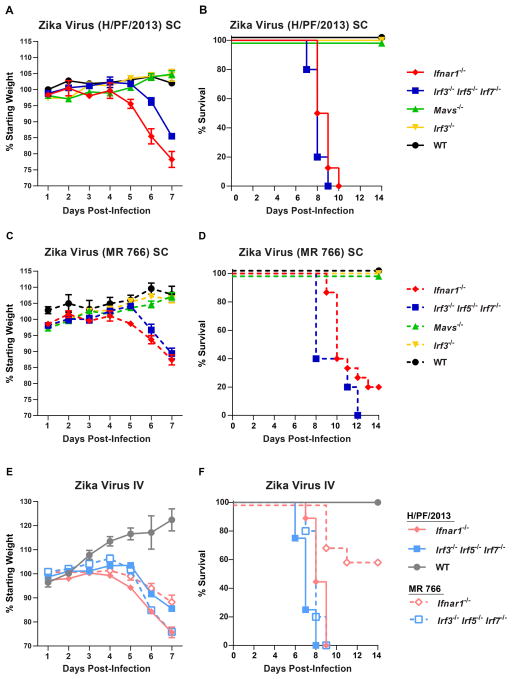
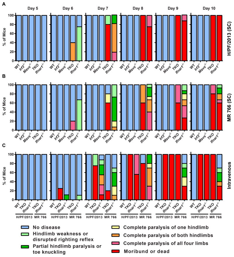
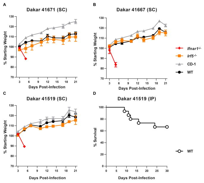
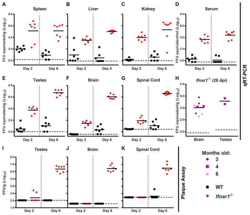
The ongoing Zika virus (ZIKV) epidemic and unexpected clinical outcomes, including Guillain-Barré syndrome and birth defects, has brought an urgent need for animal models. We evaluated infection and pathogenesis with contemporary and historical ZIKV strains in immunocompetent mice and mice lacking components of the antiviral response. Four- to six-week-old Irf3(-/-)Irf5(-/-)Irf7(-/-) triple knockout mice, which produce little interferon α/β, and mice lacking the interferon receptor (Ifnar1(-/-)) developed neurological disease and succumbed to ZIKV infection, whereas single Irf3(-/-), Irf5(-/-), and Mavs(-/-) knockout mice exhibited no overt illness. Ifnar1(-/-) mice sustained high viral loads in the brain and spinal cord, consistent with evidence that ZIKV causes neurodevelopmental defects in human fetuses. The testes of Ifnar1(-/-) mice had the highest viral loads, which is relevant to sexual transmission of ZIKV. This model of ZIKV pathogenesis will be valuable for evaluating vaccines and therapeutics as well as understanding disease pathogenesis.


Figures

Figure 1
Figure 1. Mice lacking IFN-α/β responses are susceptible to ZIKV disease
5 to 6 week-old mice of the indicated genotypes were inoculated with 102 FFU of ZIKV strain H/PF/2013 or MR 766 by a subcutaneous route (SC) in the footpad (AD) or by an intravenous route (IV) (E–F). Mice were weighed daily and weights are expressed as percentage of body weight prior to infection (A, C, and E). Results shown are the mean ± standard error of the mean (SEM) of the indicated number of mice per group. Data are censored at 7 days after infection, as mice in some groups died. Lethality was monitored for 14 days (B, D, and F). A and B: n = 16 (Ifnar1−/−), 5 (Irf3−/− Irf5−/− Irf7−/− TKO, Mavs−/−, WT), 9 (Irf3−/−). C and D: n = 15 (Ifnar1−/−), 5 (Irf3−/− Irf5−/− Irf7−/− TKO, Mavs−/−), 9 (Irf3−/−), 3 (WT). E and F: n = 9 (Ifnar1−/− H/PF/2013), 4 (Irf3−/− Irf5−/− Irf7−/− TKO, WT H/PF/2013), 10 (Ifnar1−/− MR 766), 5 (Irf3−/− Irf5−/− Irf7−/− TKO MR 766).
Figure 2
Figure 2. ZIKV infection produces neurologic disease in Ifnar1−/− and Irf3−/− Irf5−/− Irf7−/− TKO mice
Mice of the indicated genotypes were infected with ZIKV strain H/PF/2013 (A) or MR 766 (B) by a subcutaneous (SC) (A and B) or intravenous (C) route and disease signs were assessed daily for 10 days. The percentage of each group of mice displaying the indicated signs is shown. These are the same mice evaluated for weight loss and lethality in Figure 1.
Figure 3
Figure 3. ZIKV strains from Senegal cause lethal disease in 4 week-old Ifnar1−/− mice and 1 week-old WT mice
A–C. 4 week-old WT (C57BL/6), CD-1, Irf5−/−, and Ifnar1−/− mice were infected with 103 FFU of one of three ZIKV strains isolated from mosquitoes in Senegal (Dakar 41671, 41667, or 41519) by a subcutaneous route (SC). Weights were obtained over 21 days and are expressed as a percentage of starting weight. Ifnar1−/− mice succumbed to the infection after 6 days. Results shown are the mean ± SEM of 4 to 5 mice per group (WT, CD-1, and Irf5−/−) or 2 to 3 mice per group (Ifnar1−/−). D. 1 week-old WT mice (n = 15) were infected with 104 FFU of ZIKV Dakar 41519 by intraperitoneal (IP) injection and lethality was monitored for 30 days.
Figure 4
Figure 4. Older Ifnar1−/− mice remain susceptible to ZIKV infection
3, 4, or 6 month-old Ifnar1−/− C57BL/6 mice were infected with 103 FFU of ZIKV (H/PF/2013) by a subcutaneous route. A. Mice were weighed over 21 days with data censored at 9 days after infection, when mice began to succumb. Weights are expressed as a percentage of starting weight, shown as the mean ± SEM of 5 mice per group. B. Survival was monitored for 21 days.
Figure 5
Figure 5. Treatment with an IFNAR1 blocking MAb increases ZIKV viremia in WT mice
An IFNAR1-blocking MAb (MAR1-5A3) or isotype control MAb (GIR-208) was administered to 4 to 5 week-old WT C57BL/6 mice by intraperitoneal injection. Mice were infected with 103 FFU of ZIKV (H/PF/2013) by a subcutaneous route. A. ZIKV RNA in serum was measured at 3 days after infection by qRT-PCR. Data are expressed as FFU equivalents per ml after normalization to a standard curve generated in parallel. **, P < 0.01; ***, P < 0.001 compared to isotype control (Mann-Whitney test). B. Weights are expressed as a percentage of starting weight, shown as the mean ± SEM of 6 mice per group.
Figure 6
Figure 6. Tissue tropism of ZIKV in WT and Ifnar1−/− mice
4 to 6 week-old WT or Ifnar1−/− C57BL/6 male mice were inoculated with 103 FFU of ZIKV H/PF/2013 by a subcutaneous route. At day 2 or day 6 after infection, the indicated tissues were harvested, weighed, homogenized, and analyzed by qRT-PCR (A–G) or plaque assay (I–K). Results are combined from two independent experiments with a total of 7 mice per group; data are expressed as FFU equivalents per gram or ml after normalization to a standard curve generated in parallel (A–G) or as PFU per gram (I–K). H. Brain and testes were harvested 28 days after infection from 3, 4, or 6-month old Ifnar1−/− mice inoculated with 103 FFU of ZIKV H/PF/2013 and viral RNA levels were determined by qRT-PCR. These mice are the survivors from experiments shown in Figure 4 (all surviving mice for brain tissue, males only for testes).

Comment in

Similar articles

Cited by

References

    1. Alkan C, Zapata S, Bichaud L, Moureau G, Lemey P, Firth AE, Gritsun TS, Gould EA, de Lamballerie X, Depaquit J, et al. Ecuador Paraiso Escondido Virus, a New Flavivirus Isolated from New World Sand Flies in Ecuador, Is the First Representative of a Novel Clade in the Genus Flavivirus. J Virol. 2015;89:11773–11785. - PMC - PubMed
    1. Ank N, Iversen MB, Bartholdy C, Staeheli P, Hartmann R, Jensen UB, Dagnaes-Hansen F, Thomsen AR, Chen Z, Haugen H, et al. An important role for type III interferon (IFN-lambda/IL-28) in TLR-induced antiviral activity. J Immunol. 2008;180:2474–2485. - PubMed
    1. Atkinson B, Hearn P, Afrough B, Lumley S, Carter D, Aarons EJ, Simpson AJ, Brooks TJ, Hewson R. Detection of Zika Virus in Semen. Emerging infectious diseases. 2016;22 - PMC - PubMed
    1. Baronti C, Piorkowski G, Charrel RN, Boubis L, Leparc-Goffart I, de Lamballerie X. Complete coding sequence of zika virus from a French polynesia outbreak in 2013. Genome announcements. 2014;2:e00500–14. - PMC - PubMed
    1. Bell TM, Field EJ, Narang HK. Zika virus infection of the central nervous system of mice. Archiv fur die gesamte Virusforschung. 1971;35:183–193. - PubMed