

An Invariant-Based Damage Model for Human and Animal Skins
- PMID: 27066788
- PMCID: PMC5042997
- DOI: 10.1007/s10439-016-1603-9
An Invariant-Based Damage Model for Human and Animal Skins
Erratum in
-
Erratum to: An Invariant-Based Damage Model for Human and Animal Skins.Ann Biomed Eng. 2016 Oct;44(10):3123. doi: 10.1007/s10439-016-1630-6. Ann Biomed Eng. 2016. PMID: 27193015 Free PMC article. No abstract available.
Abstract
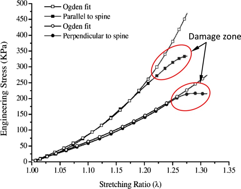
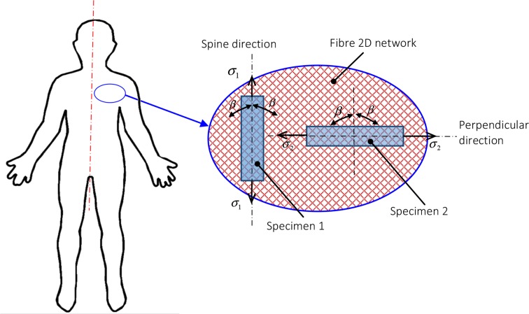
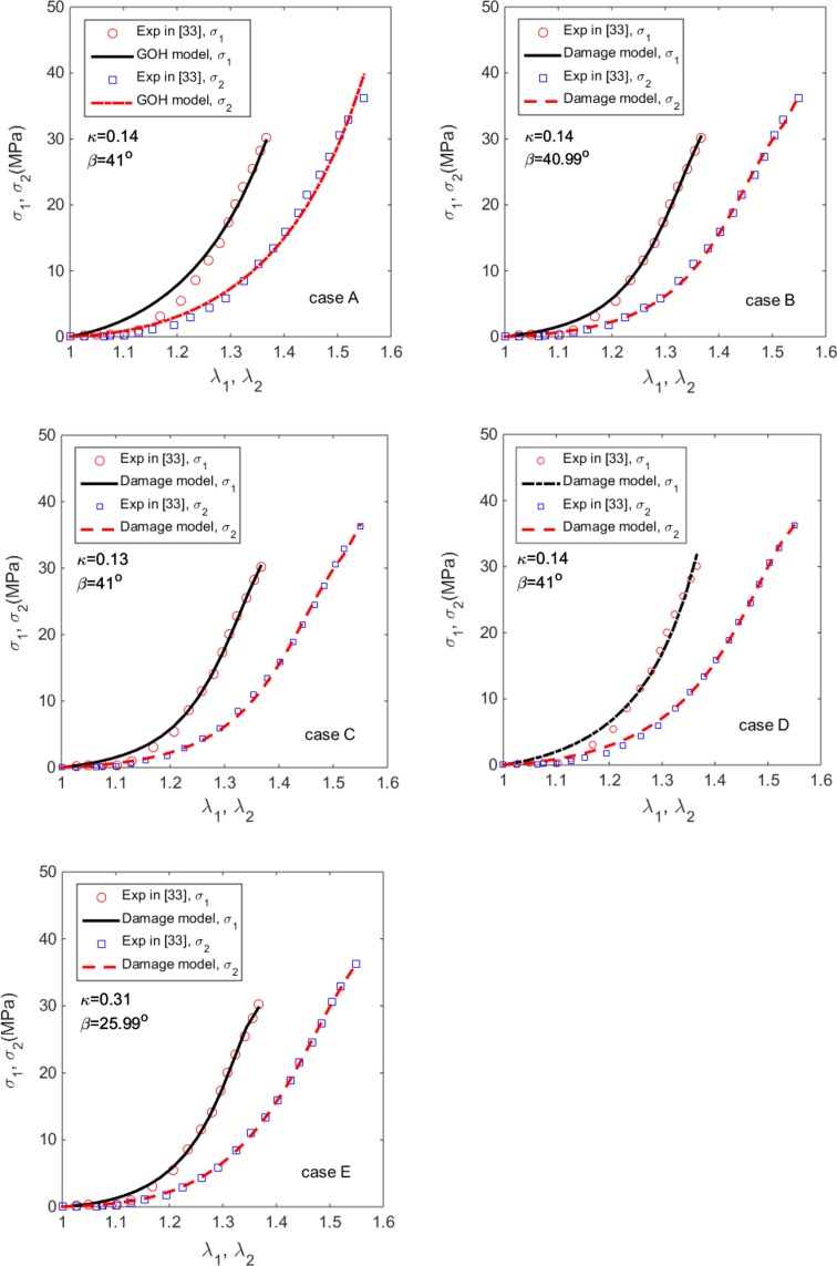
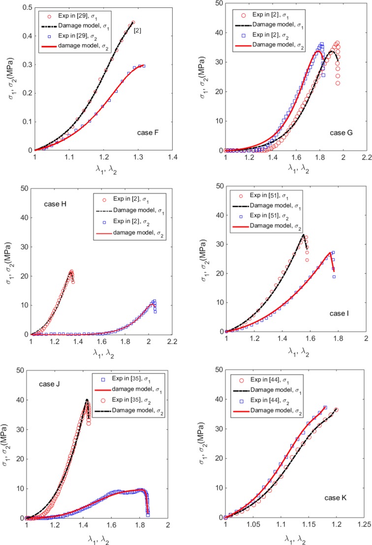
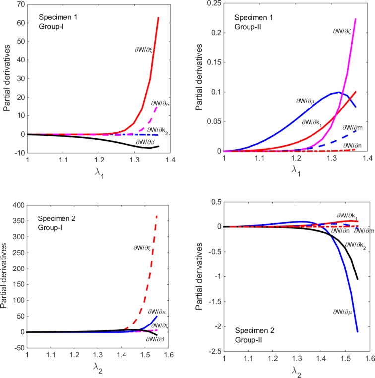
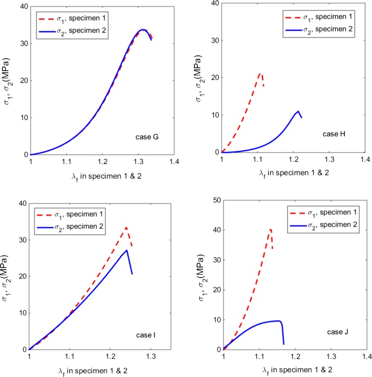
Constitutive modelling of skins that account for damage effects is important to provide insight for various clinical applications, such as skin trauma and injury, artificial skin design, skin aging, disease diagnosis, surgery, as well as comparative studies of skin biomechanics between species. In this study, a new damage model for human and animal skins is proposed for the first time. The model is nonlinear, anisotropic, invariant-based, and is based on the Gasser-Ogden-Holzapfel constitutive law initially developed for arteries. Taking account of the mean collagen fibre orientation and its dispersion, the new model can describe a wide range of skins with damage. The model is first tested on the uniaxial test data of human skin and then applied to nine groups of uniaxial test data for the human, swine, rabbit, bovine and rhino skins. The material parameters can be inversely estimated based on uniaxial tests using the optimization method in MATLAB with a root mean square error ranged between 2.15% and 12.18%. A sensitivity study confirms that the fibre orientation dispersion and the mean fibre angle are among the most important factors that influence the behaviour of the damage model. In addition, these two parameters can only be reliably estimated if some histological information is provided. We also found that depending on the location of skins, the tissue damage may be brittle controlled by the fibre breaking limit (i.e., when the fibre stretch is greater than 1.13-1.32, depending on the species), or ductile (due to both the fibre and the matrix damages). The brittle damages seem to occur mostly in the back, and the ductile damages are seen from samples taken from the belly. The proposed constitutive model may be applied to various clinical applications that require knowledge of the mechanical response of human and animal skins.
Keywords: Constitutive model; Damage; Fibre orientation; Fibre orientation dispersion; Inverse problem; Skin.
Figures
Similar articles
-
On the AIC-based model reduction for the general Holzapfel-Ogden myocardial constitutive law.Biomech Model Mechanobiol. 2019 Aug;18(4):1213-1232. doi: 10.1007/s10237-019-01140-6. Epub 2019 Apr 3. Biomech Model Mechanobiol. 2019. PMID: 30945052 Free PMC article.
-
Automated estimation of collagen fibre dispersion in the dermis and its contribution to the anisotropic behaviour of skin.Ann Biomed Eng. 2012 Aug;40(8):1666-78. doi: 10.1007/s10439-012-0542-3. Epub 2012 Mar 17. Ann Biomed Eng. 2012. PMID: 22427196
-
Unravelling the mechanics of gastric tissue: A comparison of constitutive models, damage probability and microstructural insights.J Mech Behav Biomed Mater. 2024 Dec;160:106712. doi: 10.1016/j.jmbbm.2024.106712. Epub 2024 Sep 3. J Mech Behav Biomed Mater. 2024. PMID: 39276436
-
Power type strain energy function model and prediction of the anisotropic mechanical properties of skin using uniaxial extension data.Med Biol Eng Comput. 2013 Oct;51(10):1147-56. doi: 10.1007/s11517-013-1098-6. Epub 2013 Jul 18. Med Biol Eng Comput. 2013. PMID: 23864550
-
Hyperelastic modelling of arterial layers with distributed collagen fibre orientations.J R Soc Interface. 2006 Feb 22;3(6):15-35. doi: 10.1098/rsif.2005.0073. J R Soc Interface. 2006. PMID: 16849214 Free PMC article. Review.
Cited by
-
Mathematical and computational modelling of skin biophysics: a review.Proc Math Phys Eng Sci. 2017 Jul;473(2203):20170257. doi: 10.1098/rspa.2017.0257. Epub 2017 Jul 26. Proc Math Phys Eng Sci. 2017. PMID: 28804267 Free PMC article. Review.
-
Constitutive law of healthy gallbladder walls in passive state with damage effect.Biomed Eng Lett. 2019 Feb 6;9(2):189-201. doi: 10.1007/s13534-019-00098-9. eCollection 2019 May. Biomed Eng Lett. 2019. PMID: 31168424 Free PMC article.
-
A Review on Damage and Rupture Modelling for Soft Tissues.Bioengineering (Basel). 2022 Jan 10;9(1):26. doi: 10.3390/bioengineering9010026. Bioengineering (Basel). 2022. PMID: 35049735 Free PMC article. Review.
References
Publication types
MeSH terms
LinkOut - more resources
Full Text Sources
Other Literature Sources
Medical
