

Evaluation of right ventricular function in fetal hypoplastic left heart syndrome using spatio-temporal image correlation (STIC)
- PMID: 27066831
- PMCID: PMC4827237
- DOI: 10.1186/s12947-016-0056-5
Evaluation of right ventricular function in fetal hypoplastic left heart syndrome using spatio-temporal image correlation (STIC)
Abstract
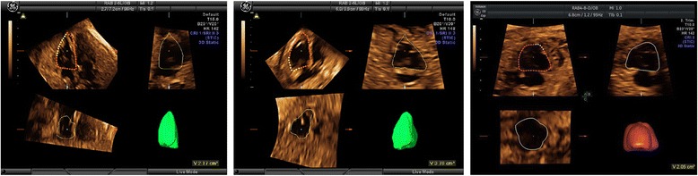
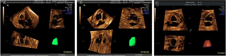
Background: Postnatal outcome of fetuses with hypoplastic left heart syndrome (HLHS) is mainly determined by right ventricular function. In the present study we used spatio-temporal image correlation (STIC) to assess right ventricular function of fetuses with HLHS.
Methods: Three-dimensional ultrasound with STIC technique was used to acquire heart images from fetuses that had HLHS and the normal controls, between 24(+0) and 37(+6) weeks of gestation. Right ventricular end-diastolic volume (RVEDV) and right ventricular end-systolic volume (RVESV) were determined using the virtual organ computer-aided analysis software, and the parameters of right ventricular function were calculated.
Results: Both RVEDV and RVESV were found to be significantly higher in fetuses with HLHS as compared to that in normal controls (P < 0.001). There were no significant differences in the parameters between fetuses with and without a visible left ventricular cavity (P > 0.05). Compared to fetuses with HLHS plus mild tricuspid regurgitation (TR), fetuses with HLHS plus severe TR exhibited lower right ventricular stroke volume (RVSV), right ventricular cardiac output (RVCO) and standardized RVCO (P < 0.05). The right ventricular ejection fraction (RVEF) was significantly lower in HLHS fetuses that had severe TR (P < 0.001).
Conclusion: As the right ventricle is solely responsible for maintenance of circulation, the right ventricular systolic function undergoes compensatory enhancement in fetuses with HLHS and mild TR, compared to that in normal controls. Size of the left ventricle does not significantly affect the right ventricular function in HLHS. However, right ventricular systolic function may be impaired prenatally in HLHS fetuses that have severe TR.
Keywords: Fetal hypoplastic left heart syndrome; Right ventricular function; Spatio-temporal image correlation.
Figures
Similar articles
-
Right Ventricular Systolic-to-Diastolic Time Index: Hypoplastic Left Heart Fetuses Differ Significantly from Normal Fetuses.J Am Soc Echocardiogr. 2016 Feb;29(2):143-9. doi: 10.1016/j.echo.2015.08.014. Epub 2015 Sep 26. J Am Soc Echocardiogr. 2016. PMID: 26394829
-
Right ventricular function in fetal hypoplastic left heart syndrome.J Am Soc Echocardiogr. 2012 Oct;25(10):1068-74. doi: 10.1016/j.echo.2012.06.005. Epub 2012 Jul 12. J Am Soc Echocardiogr. 2012. PMID: 22795200
-
Evaluation of right ventricular function in fetuses with hypoplastic left heart syndrome using tissue Doppler techniques.Ultrasound Obstet Gynecol. 2015 Jun;45(6):670-7. doi: 10.1002/uog.14736. Epub 2015 May 11. Ultrasound Obstet Gynecol. 2015. PMID: 25418127
-
Role of cardiac magnetic resonance strain analysis in patients with hypoplastic left heart syndrome in evaluating right ventricular (dys)function: a systematic review.Eur J Cardiothorac Surg. 2021 Sep 11;60(3):497-505. doi: 10.1093/ejcts/ezab105. Eur J Cardiothorac Surg. 2021. PMID: 34331061
-
Assessment of the right ventricle by echocardiography: a primer for cardiac sonographers.J Am Soc Echocardiogr. 2009 Jul;22(7):776-92; quiz 861-2. doi: 10.1016/j.echo.2009.04.027. J Am Soc Echocardiogr. 2009. PMID: 19560657 Review.
Cited by
-
Evaluation of right ventricular volume and systolic function in normal fetuses using intelligent spatiotemporal image correlation.World J Clin Cases. 2019 Aug 6;7(15):2003-2012. doi: 10.12998/wjcc.v7.i15.2003. World J Clin Cases. 2019. PMID: 31423432 Free PMC article.
-
Evaluation of Right Ventricular Function and Myocardial Microstructure in Fetal Hypoplastic Left Heart Syndrome.J Clin Med. 2022 Jul 30;11(15):4456. doi: 10.3390/jcm11154456. J Clin Med. 2022. PMID: 35956075 Free PMC article.
-
Fetal cardiac magnetic resonance imaging of the descending aorta in suspected left-sided cardiac obstructions.Front Cardiovasc Med. 2023 Dec 1;10:1285391. doi: 10.3389/fcvm.2023.1285391. eCollection 2023. Front Cardiovasc Med. 2023. PMID: 38107261 Free PMC article.
References
Publication types
MeSH terms
LinkOut - more resources
Full Text Sources
Other Literature Sources
Research Materials
