. 2016 Apr 12:7:11303.
doi: 10.1038/ncomms11303.

A mutation in the atrial-specific myosin light chain gene (MYL4) causes familial atrial fibrillation

Affiliations

A mutation in the atrial-specific myosin light chain gene (MYL4) causes familial atrial fibrillation

Nathan Orr et al. Nat Commun. .

Abstract

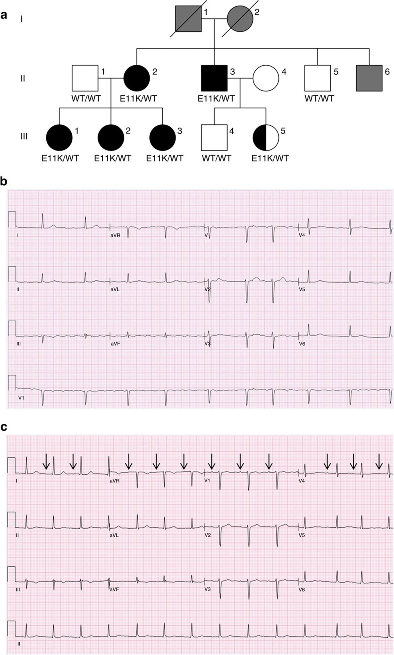
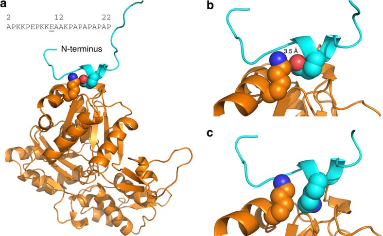
Atrial fibrillation (AF), the most common arrhythmia, is a growing epidemic with substantial morbidity and economic burden. Mechanisms underlying vulnerability to AF remain poorly understood, which contributes to the current lack of highly effective therapies. Recognizing mechanistic subtypes of AF may guide an individualized approach to patient management. Here, we describe a family with a previously unreported syndrome characterized by early-onset AF (age <35 years), conduction disease and signs of a primary atrial myopathy. Phenotypic penetrance was complete in all mutation carriers, although complete disease expressivity appears to be age-dependent. We show that this syndrome is caused by a novel, heterozygous p.Glu11Lys mutation in the atrial-specific myosin light chain gene MYL4. In zebrafish, mutant MYL4 leads to disruption of sarcomeric structure, atrial enlargement and electrical abnormalities associated with human AF. These findings describe the cause of a rare subtype of AF due to a primary, atrial-specific sarcomeric defect.


Figures

Figure 1
Figure 1. Kindred with familial atrial fibrillation caused by mutation of MYL4.
(a) Kindred structure. Affected members with AF are denoted by black filled symbols; subject III.5 is half-filled, reflecting a partial phenotype. Family members with unknown phenotype are filled grey. Protein alterations encoded at the MYL4 locus are indicated. (b) Electrocardiogram of index case (subject III.3) demonstrating atrial fibrillation with slow ventricular rate response while off medication. (c) Electrocardiogram of sister of the index case (subject III.2) during normal sinus rhythm. Arrows point to very low amplitude P-waves. Prolonged PR interval (atrial–ventricular conduction time) is also present.
Figure 2
Figure 2. Electrocardiograms and whole hearts from WT transgenic and E17K transgenic zebrafish.
(a) WT transgenics exhibit a regular heart rate with electrocardiogram parameters typical for adult zebrafish. E17K transgenics display significantly slower sino-atrial activity and increased heart rate irregularity compared with controls. (b) Signal-averaged P-wave duration is prolonged in E17K transgenics, indicative of slower atrial conduction time. (c) Atrial area has been traced in isolated whole-WT transgenic and -E17K transgenic hearts (dotted lines), showing enlarged atrial chamber size in E17K transgenic.
Figure 3
Figure 3. Myofibrillar organization and sarcomeric structure in WT and E17K transgenics.
(a) A WT transgenic atrium at larval stage (5 d.p.f.) in the Tg(myl7:LifeAct-GFP)s974 background shows GFP-labelled F-actin, with myofibrils organized linearly across the atrium, either up-and-down or left-to-right. The myofibrils have a ‘dotted line' pattern, with well-demarcated box-like shapes indicating normal arrangement of F-actin in the myofibrils. The non-fluorescent gaps (arrows) represent the actin-poor H-zones. (b) In an E17K transgenic larval atrium, some myofibrils appear unaffected (arrows), but there are large areas where myofibrils are not linearly organized and actin localization appears abnormal, giving a stippled appearance to the sarcomeres without clear H-zones (arrowheads). (c) Electron microscopy of adult WT zebrafish atrium shows clear H-zones and Z-disks. (d) Electron microscopy of E17K transgenic atrium shows preserved H-zones but absent Z-disks (arrows). H, H-zone. Z, Z-disk.
Figure 4
Figure 4. Secondary structure prediction for Myl4 WT and mutant N-termini.
(a) In the presence of WT E11, a type II β-turn is predicted with the formation of a stable hydrogen bond between E11 and K14. (b) Substitution of positively charged K11 for E11 results in loss of the predicted β-turn. C, coil; T, β-turn.
Figure 5
Figure 5. Predicted interaction of Myl4 with putative actin-binding site.
(a) WT E11–Myl4 is in close proximity to K358 of actin and is predicted to form a stable electrostatic interaction between the negatively charged oxygen of E11 and positively charged nitrogen of actin K358 (b). (c) This electrostatic interaction within the actin-binding site is lost with substitution of positively charged K11. Dark-blue sphere, positively charged nitrogen molecule. Red sphere, negatively charged oxygen molecule.

References

    1. Ball J., Carrington M. J., McMurray J. J. & Stewart S. Atrial fibrillation: profile and burden of an evolving epidemic in the 21st century. Int. J. Cardiol. 167, 1807–1824 (2013). - PubMed
    1. Patel N. J. et al.. Contemporary trends of hospitalization for atrial fibrillation in the United States, 2000 through 2010: implications for healthcare planning. Circulation 129, 2371–2379 (2014). - PubMed
    1. Sorgente A., Tung P., Wylie J. & Josephson M. E. Six year follow-up after catheter ablation of atrial fibrillation: a palliation more than a true cure. Am. J. Cardiol. 109, 1179–1186 (2012). - PubMed
    1. Verma A. et al.. Discerning the incidence of symptomatic and asymptomatic episodes of atrial fibrillation before and after catheter ablation (DISCERN AF): a prospective, multicenter study. JAMA Intern. Med. 173, 149–156 (2013). - PubMed
    1. Marsman R. F., Tan H. L. & Bezzina C. R. Genetics of sudden cardiac death caused by ventricular arrhythmias. Nat. Rev. Cardiol. 11, 96–111 (2014). - PubMed

Publication types

MeSH terms