Review
. 2016 Jun;32(6):334-347.
doi: 10.1016/j.tig.2016.03.004. Epub 2016 Apr 8.

To Be Specific or Not: The Critical Relationship Between Hox And TALE Proteins

Affiliations
Review

To Be Specific or Not: The Critical Relationship Between Hox And TALE Proteins

Samir Merabet et al. Trends Genet. 2016 Jun.

Abstract

Hox proteins are key regulatory transcription factors that act in different tissues of the embryo to provide specific spatial and temporal coordinates to each cell. These patterning functions often depend on the presence of the TALE-homeodomain class cofactors, which form cooperative DNA-binding complexes with all Hox proteins. How this family of cofactors contributes to the highly diverse and specific functions of Hox proteins in vivo remains an important unsolved question. We review here the most recent advances in understanding the molecular mechanisms underlying Hox-TALE function. In particular, we discuss the role of DNA shape, DNA-binding affinity, and protein-protein interaction flexibility in dictating Hox-TALE specificity. We propose several models to explain how these mechanisms are integrated with each other in the context of the many distinct functions that Hox and TALE factors carry out in vivo.

Keywords: DNA-binding specificity; Hox proteins; SELEX-seq; SPIMs; TALE cofactors; homeodomains.


Figures

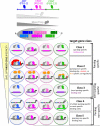
Figure 1
Figure 1. Hox and TALE members in vertebrates and invertebrates
(a). Evolution of Hox and TALE genes in Bilateria (B). Representative species for Deuterostomes (D) and for Ecdysozoa (E) and Lophotrochoza (L) branches from Protostomes (P) are Mus musculus, Drosophila melanogaster and Caenorhabditis elegans, respectively. Hox genes are organized into anterior (ant, blue-graded boxes), central (cent, pink-graded boxes) and posterior (post, green-graded boxes) paralog groups. This representation does not reflect the genomic cluster organisation. Independent duplications are indicated for central and posterior Hox genes (brackets). Boxes representing bicoid (bcd), zerknült (zen) and fushi-tarazu (ftz) are not colour-filled because these three genes strongly diverged in Drosophila. Note that PREP was specifically lost in Drosophila among Ecdysozoan species. (b). Schematic representation of motifs and domains involved in the Hox-TALE partnership. Hox proteins can use a generic (W-containing) motif and/or a specific PBC interaction motif (SPIM) to interact with the homeodomain (HD) of PBC. PBC-A and PBC-B domains interact with the MEIS-A and MEIS-B domains of Meis or Prep. Note that these complexes could also form with HD-less isoforms of Hth and Meis.
Figure 2
Figure 2. Key Figure
Different classes of target genes underlie Hox function in vivo. Target genes are classified (class 1 to 5) according to their level of specificity for Hox paralogs. Different classes of target genes contain Hox-TALE binding sites with various degrees of specificity, as indicated. Note that the level of binding site specificity does not necessarily correlate with the paralog specificity of the target gene. TALE-independent Hox target genes could also fall into those categories, as discussed elsewhere [7]. Known examples are described for class 1 (with specific activation:[–23]), class 2 (lower row, with specific repression: [24]), class 3 (with activated or repressed target genes [–15]) and class 5 (corresponding to artificial constructs: [12,19]). Target genes for class 2 (upper row, corresponding to the tritocerebrum context [18]) and class 4 are speculative (see also Figure 4a). Schematic expression profile of Hox and TALE proteins along the anterior-posterior (AP) axis of the Drosophila embryo is provided above the model. Differences in TALE expression levels might influence Hox function (see also Figure 4b). The color code for Hox paralogs and TALE cofactors is the same as in Figure 1. This color code is also used for binding sites that are paralog-specific or semi-paralog-specific. Non-paralog-specific binding sites are depicted in dark gray. Presence or absence of arrows, respectively, indicates regulation or not by the Hox-cofactor complex. Red and orange proteins in class 2 target genes highlight additional cofactors that provide cell-,(upper row) or Hox-specific (lower row) regulatory activity.
Figure 3
Figure 3. Specificity is inversely correlated to binding affinity
(a). Interaction with TALE cofactors allows Hox proteins from different paralog groups (highlighted in pink and blue) to preferentially bind to distinct sequences. In the absence of TALE factors, two Hox proteins bind to very similar sequences (left graph) while in the presence of TALE factors the same two Hox proteins exhibit distinct preferences (right graph). Representative nucleotide sequences recognized by monomers or Hox/TALE complexes are provided according to [6,27]. (b). The graph shows a hypothetical trade-off between specificity and affinity. Representative nucleotide sequences recognized by a Hox-TALE complex are provided according to [27]. High specificity and low affinity binding sites depend on both DNA shape and base recognition mechanisms as discussed in the main text. (c). Three dimensional structures illustrating the recognition of a paralog non-specific binding site (fkhcon [19], left, corresponding to case (1) in Figure 3b) and of a paralog-specific binding site (fkh [19], right, corresponding to case (2) in Figure 3b) bound by the Hox protein Scr with its cofactor Exd. Note that complex formation on the paralog-specific site involves additional Scr-specific residues (Arg3 and His-12) and the recognition of a narrow minor grove (indicated by the double black arrow). The equivalent region of the paralog-non-specific complex has a wider minor groove. Adapted from [20] and the coordinates are from Protein Data Base (PDB) structures 2r5y and 2r5z, respectively.
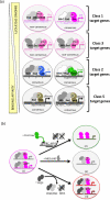
Figure 4
Figure 4. Various combinatorial relationships underlie the function of Hox-TALE complexes
(a) Examples of binding sites characterized in the cis-regulatory regions of Hox-TALE target genes. Hox-TALE binding sites that are specific or have intermediate specificity contain a consensus (e.g. forkhead (fkh, [19]) target gene) or non-consensus (e.g. rhomboid (rho, [22]), Distalless (Dll, [12]) and shavenbaby (svb, [14]) target genes) binding sites. Non-paralog specific Hox-TALE binding sites generally include a consensus binding site (e.g. Foxp1 [24], Dllcon [12] and fkhcon [19]). Binding site specificity is illustrated by the color code (dark gray is non specific). Binding site affinity is illustrated by a variable line width. The brown color of the Hox protein in class 5 target genes illustrates the ability of all paralogs to regulate a non-paralog specific enhancer. Motifs used for complex assembly on these different types of binding sites are indicated (W-containing or SPIM), as discussed in the main text. These motifs are speculative for class 2 target genes (b). Variation in the composition of Hox or TALE proteins can influence the interaction mode and, as a result, the activity of Hox-TALE complexes. An example is provided for a central Hox protein (pink) that could use either a W- motif or a SPIM to interact with the PBC and Meis cofactors in cell context (1). Three different additional cell contexts are proposed, based on published data. In cell context (2), the presence of a posterior Hox protein blocks the expression of TALE- encoding genes [16]. As a result, central Hox proteins will only regulate target genes that do not depend on TALE input. In cell context (3), the presence of a HD-less isoform in place of full length Meis does not allow the use of a SPIM for complex assembly [47]. As a consequence, the trimeric complex is not able to regulate paralog-specific target genes that depend on SPIM-mediated conformation modes. In cell context (4), the presence of Prep induces a degradation of Meis, indirectly through the titration of PBC [61], leading to the formation of Hox-PBC-Prep in place of Hox-PBC-Meis complexes. These complexes have the potential to assemble by using the W-containing motif and/or SPIMs but could potentially regulate distinct target genes compared to those regulated by Hox-PBC-Meis complexes.

References

    1. Pick L, Heffer A. Hox gene evolution: Multiple mechanisms contributing to evolutionary novelties. Ann. N. Y. Acad. Sci. 2012;1256:15–32. - PubMed
    1. Hombría JC-G, Lovegrove B. Beyond homeosis--HOX function in morphogenesis and organogenesis. Differentiation. 2003;71:461–76. - PubMed
    1. Seifert A, et al. Role of Hox genes in stem cell differentiation. World J. Stem Cells. 2015;7:583–95. - PMC - PubMed
    1. Holland PWH. Evolution of homeobox genes. Wiley Interdisciplinary Reviews: Developmental Biology. 2013;2:31–45. - PubMed
    1. Merabet S, et al. Classification of sequence signatures: a guide to Hox protein function. Bioessays. 2009;31:500–511. - PubMed

MeSH terms

LinkOut - more resources