

Biomarkers for the detection of necroptosis
- PMID: 27066893
- PMCID: PMC11108390
- DOI: 10.1007/s00018-016-2192-3
Biomarkers for the detection of necroptosis
Abstract
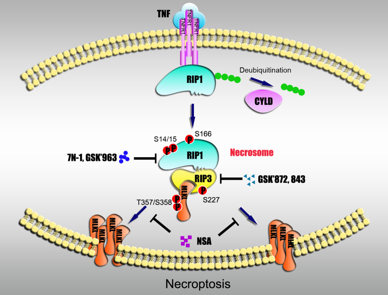
Necroptosis has been extensively studied recently, and the receptor-interacting kinase 3 (RIP3 or RIPK3) and its substrate, the pseudokinase mixed lineage kinase domain-like protein, have been discovered to be core components of this process. Classical necroptosis requires RIP1 (or RIPK1) for the activation of RIP3 through the induction of RIP1/RIP3 necrosomes. Increasing evidence from genetic and pharmacological studies has been expanding the view that necroptosis plays important roles in the etiology and/or progression of many human diseases, such as pancreatitis, ischemic injury, and neurodegenerative diseases, among others. Ongoing progress in translational research about necroptosis has highlighted the increasingly important need for the identification of biomarkers for use in disease diagnosis, monitoring, and drug development. This review presents a discussion of the current status of biomarkers that can be used to detect necroptosis both in vitro and in vivo.
Keywords: Biomarker; MLKL; Necroptosis; RIP1; RIP3; Regulated necrosis.
Figures
Similar articles
-
RIP1, RIP3, and MLKL Contribute to Cell Death Caused by Clostridium perfringens Enterotoxin.mBio. 2019 Dec 17;10(6):e02985-19. doi: 10.1128/mBio.02985-19. mBio. 2019. PMID: 31848291 Free PMC article.
-
PI3K mediates tumor necrosis factor induced-necroptosis through initiating RIP1-RIP3-MLKL signaling pathway activation.Cytokine. 2020 May;129:155046. doi: 10.1016/j.cyto.2020.155046. Epub 2020 Feb 28. Cytokine. 2020. PMID: 32114297
-
Distinct roles of RIP1-RIP3 hetero- and RIP3-RIP3 homo-interaction in mediating necroptosis.Cell Death Differ. 2014 Nov;21(11):1709-20. doi: 10.1038/cdd.2014.77. Epub 2014 Jun 6. Cell Death Differ. 2014. PMID: 24902902 Free PMC article.
-
Necrosome core machinery: MLKL.Cell Mol Life Sci. 2016 Jun;73(11-12):2153-63. doi: 10.1007/s00018-016-2190-5. Epub 2016 Apr 5. Cell Mol Life Sci. 2016. PMID: 27048809 Free PMC article. Review.
-
Necroptosis in health and diseases.Semin Cell Dev Biol. 2014 Nov;35:14-23. doi: 10.1016/j.semcdb.2014.07.013. Epub 2014 Aug 1. Semin Cell Dev Biol. 2014. PMID: 25087983 Review.
Cited by
-
CHIP ameliorates cerebral ischemia-reperfusion injury by attenuating necroptosis and inflammation.Aging (Albany NY). 2021 Dec 14;13(23):25564-25577. doi: 10.18632/aging.203774. Epub 2021 Dec 14. Aging (Albany NY). 2021. PMID: 34905731 Free PMC article.
-
Vitexicarpin Induces Apoptosis and Inhibits Metastatic Properties via the AKT-PRAS40 Pathway in Human Osteosarcoma.Int J Mol Sci. 2024 Mar 22;25(7):3582. doi: 10.3390/ijms25073582. Int J Mol Sci. 2024. PMID: 38612399 Free PMC article.
-
Drug discovery and formulation development for acute pancreatitis.Drug Deliv. 2020 Dec;27(1):1562-1580. doi: 10.1080/10717544.2020.1840665. Drug Deliv. 2020. PMID: 33118404 Free PMC article. Review.
-
Gentiopicroside-Induced gastric cancer necroptosis via the HIF-1 signaling pathway: A study involving molecular docking and experimental validation.PLoS One. 2024 Nov 21;19(11):e0311152. doi: 10.1371/journal.pone.0311152. eCollection 2024. PLoS One. 2024. PMID: 39570876 Free PMC article.
-
Latifolin, a Natural Flavonoid, Isolated from the Heartwood of Dalbergia odorifera Induces Bioactivities through Apoptosis, Autophagy, and Necroptosis in Human Oral Squamous Cell Carcinoma.Int J Mol Sci. 2022 Nov 7;23(21):13629. doi: 10.3390/ijms232113629. Int J Mol Sci. 2022. PMID: 36362414 Free PMC article.
References
Publication types
MeSH terms
Substances
LinkOut - more resources
Full Text Sources
Other Literature Sources
Medical
Miscellaneous
