. 2016 Apr 11:16:10.
doi: 10.1186/s40644-016-0065-5.

How to use CT texture analysis for prognostication of non-small cell lung cancer

Affiliations

How to use CT texture analysis for prognostication of non-small cell lung cancer

Kenneth A Miles. Cancer Imaging. .

Abstract

Patients with non-small cell lung cancer frequently demonstrate differing clinical courses, even when they express the same tumour stage. Additional markers of prognostic significance could allow further stratification of treatment for these patients. By generating quantitative information about tumour heterogeneity as reflected by the distribution of pixel values within the tumour, CT texture analysis (CTTA) can provide prognostic information for patients with NSCLC. In addition to describing the practical application of CTTA to NSCLC, this article discusses a range of issues that need to be addressed when CTTA is included as part of routine clinical care as opposed to its use in a research setting. The use of quantitative imaging to provide prognostic information is a new and exciting development within cancer imaging that can expand the imaging specialist's existing role in tumour evaluation. Derivation of prognostic information through the application of image processing techniques such as CTTA, to images acquired as part of routine care can help imaging specialists make best use of the technologies they deploy for the benefit of patients with cancer.


Figures

Fig. 1
Fig. 1
The potential for prognostic biomarkers to stratify care for patients with NSCLC. CT showing left upper lobe NSCLC at initial staging a. Based on current practice, the patient underwent surgery without adjuvant chemotherapy. CT performed 25 months later shows local recurrence b. A biomarker deployed at staging may have categorised the patient as high-risk for recurrence, implying a potential benefit from adjuvant chemotherapy
Fig. 2
Fig. 2
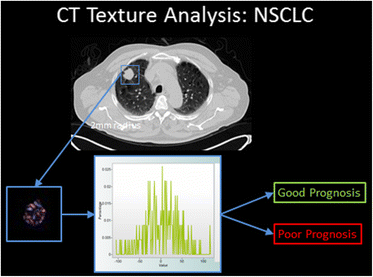
Summary of the filtration-histogram method for CTTA. The conventional CT image (top) is filtered to highlight objects of a pre-selected size. The distribution of tumour features within the filtered image as assessed using standard statistical parameters derived from the corresponding histogram, provides an indication of prognosis
Fig. 3
Fig. 3
When using automatic segmentation for tumour Regions of Interest (ROIs), the initial manually constructed ROI (a) can include surrounding lung. The segmentation software then redifines the ROI to exclude lung tissue b. The filtered tumour image (c) is used for derivation of texture parameters by histogram analysis
Fig. 4
Fig. 4
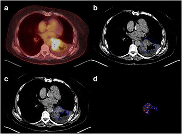
Left lower lobe NSCLC showing cavitation and adjacent consolidation. The fused FDG-PET/CT image (a) and narrow CT windows (b) can assist identification of the tumour margins. Using automatic segmentation, the initial manually constructed ROI (b) includes adjacent lung and the area of cavitation but excludes the adjacent mediastinal structures and pulmonary consolidation. The final ROI defined by the automated segmentation procedure (c) exlcudes the adjacent lung and area of cavitation. The final filtered tumour image is shown in (d)
Fig. 5
Fig. 5
Right lower lobe NSCLC showing necrosis without cavitation (photopaenia on FDG-PET/CT) and adjacent pulmonary consolidation (a). Due to minimal contact with aerated lung, the tumour ROI has been constructed manually (b), using the fused PET/CT image and narrow windows for guidance. The area of necrosis without cavitation is included in the the ROI and the final filtered tumour image (c)
Fig. 6
Fig. 6
MAPK pathway and CTTA in NSCLC. Dotted lines indicate correlations between MAPK biology and CTTA in NSCLC demonstrated through clinical research

References

    1. Win T, Miles KA, Janes SM, Ganeshan B, Shastry M, Endozo R, Meagher M, Shortman RI, Wan S, Kayani I, Ell PJ, Groves AM. Tumor heterogeneity and permeability as measured on the CT component of PET/CT predict survival in patients with Non-Small Cell Lung Cancer. Clin Cancer Res. 2013;19:3591–9. doi: 10.1158/1078-0432.CCR-12-1307. - DOI - PubMed
    1. Fried DV, Tucker SL, Zhou S, Liao Z, Mawlawi O, Ibbott G, Court LE. Prognostic value and reproducibility of pretreatment CT texture features in stage III Non-Small Cell Lung Cancer. Int J Radiation Oncol Biol Phys. 2014;90:834–842. doi: 10.1016/j.ijrobp.2014.07.020. - DOI - PMC - PubMed
    1. Weiss GJ, Ganeshan B, Miles KA, Campbell DH, Cheung PY, Frank S, Korn RL. Non-invasive image texture analysis differentiates K-ras mutation from pan-wildtype NSCLC and is prognostic. PLoS One. 2014;9:e100244. doi: 10.1371/journal.pone.0100244. - DOI - PMC - PubMed
    1. Bluthgen MV, Caramella C, Faire L, Rosellini S, Facchinetti F, Haspinger E, Ferte C, Ammari S, Michiels S, Soria JC, Besse B. Prognostic value of texture analysis in advanced Non-Small Cell Lung Cancer (NSCLC). Europ J Cancer. 2015;51:S645–S646.
    1. Caramella C, Bluthgen MV, Rosellini S, Leduc C, Facchinetti F, Haspinger E, Ferte C, Michiels S, Soria JC, Besse B. Prognostic value of texture analysis and correlation with molecular profile in EGFR mutated/ALK rearranged advanced Non-Small Cell Lung Cancer (NSCLC). Europ J Cancer. 2015;51:S647-S648.

MeSH terms

Substances