Case Reports
. 2016 Apr 11:16:45.
doi: 10.1186/s12883-016-0566-7.

The value of early and comprehensive diagnoses in a human fetus with hydrocephalus and progressive obliteration of the aqueduct of Sylvius: Case Report

Affiliations
Case Reports

The value of early and comprehensive diagnoses in a human fetus with hydrocephalus and progressive obliteration of the aqueduct of Sylvius: Case Report

Eduardo Ortega et al. BMC Neurol. .

Abstract

Background: Mutant rodent models have highlighted the importance of the ventricular ependymal cells and the subcommissural organ (a brain gland secreting glycoproteins into the cerebrospinal fluid) in the development of fetal onset hydrocephalus. Evidence indicates that communicating and non-communicating hydrocephalus can be two sequential phases of a single pathological phenomenon triggered by ependymal disruption and/or abnormal function of the subcommissural organ. We have hypothesized that a similar phenomenon may occur in human cases with fetal onset hydrocephalus.

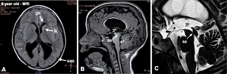
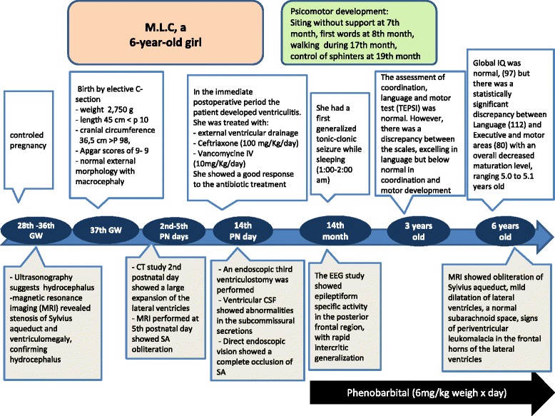
Case presentation: We report here on a case of human fetal communicating hydrocephalus with no central nervous system abnormalities other than stenosis of the aqueduct of Sylvius (SA) that became non-communicating hydrocephalus during the first postnatal week due to obliteration of the cerebral aqueduct. The case was followed closely by a team of basic and clinic investigators allowing an early diagnosis and prediction of the evolving pathophysiology. This information prompted neurosurgeons to perform a third ventriculostomy at postnatal day 14. The fetus was monitored by ultrasound, computerized axial tomography and magnetic resonance imaging (MRI). After birth, the follow up was by MRI, electroencephalography and neurological and neurocognitive assessments. Cerebrospinal fluid (CSF) collected at surgery showed abnormalities in the subcommissural organ proteins and the membrane proteins L1-neural cell adhesion molecule and aquaporin-4. The neurological and neurocognitive assessments at 3 and 6 years of age showed neurological impairments (epilepsy and cognitive deficits).

Conclusions: (1) In a hydrocephalic fetus, a stenosed SA can become obliterated at perinatal stages. (2) In the case reported, a close follow up of a communicating hydrocephalus detected in utero allowed a prompt postnatal surgery aiming to avoid as much brain damage as possible. (3) The clinical and pathological evolution of this patient supports the possibility that the progressive stenosis of the SA initiated during the embryonic period may have resulted from ependymal disruption of the cerebral aqueduct and dysfunction of the subcommissural organ. The analysis of subcommissural organ glycoproteins present in the CSF may be a valuable diagnostic tool for the pathogenesis of congenital hydrocephalus.

Keywords: Aqueduct of Sylvius; Case study; Cerebral aqueduct; Cerebrospinal fluid; Congenital hydrocephalus; Stenosis; Subcommissural organ.


Figures

Fig. 1
Fig. 1
Progressive obliteration of the aqueduct of Sylvius (SA) in hydrocephalus. a An ultrasound of the fetal patient at 36 GW demonstrating dilation of the lateral ventricles (arrows). b MRI at 33 GW. The third ventricle (3°) and posterior horns of the lateral ventricles (ph) are dilated. c MRI at 33 weeks. Stenosis of the SA is shown. d Detailed magnification of the area framed in C showing stenosis of the SA. 4°, fourth ventricle. e CT at 39 GW (or the 2nd PN day). The lateral (LV) and third (3°) ventricles are dilated. f MRI of the brain on the 5th postnatal day, sagittal T2 imaging. The SA is obliterated
Fig. 2
Fig. 2
MRI findings at 6th years of age. a MRI (transverse T1 imaging) showing mild ventricular dilatation, a normal subarachnoid space (sas), signs of periventricular leukomalacia (lk) in the frontal horns of the lateral ventricles, and a glial scar (gs) in the frontal subcortical zone. b, c MRI (sagittal T1 and sagittal T2 imaging, respectively) showing complete obliteration of the SA
Fig. 3
Fig. 3
The human subcommissural organ (SCO). a Line drawing of a human brain showing the location of the SCO (red rectangle). b Sagittal section through the epithalamus of a 32 GW human embryo (for orientation see rectangle in previous figure) showing the SCO, posterior commissure (pm), pineal gland (P), third ventricle (3°) and SA. Inset. Ependyma of a human SCO (see frame in B), immunostained for SCO-spondin. Scale bar B 350 µm, inset 50 µm. c Histograms of microdensitometric recordings of immunoblots with anti-P15, shown in (d). d Immunoblots of CSF samples from the hydrocephalic case and from a 33rd GW fetus with an arachnoid cyst, using anti-P15 and anti-hSCO antisera. Blue arrows point to compounds present in the hydrocephalic CSF and missing in the control; red arrows indicate compounds that are present in the control but missing or at lower concentration in the hydrocephalic CSF. e Histograms of microdensitometric recording of immunoblots with anti-P15, shown in (d). Control, CSF obtained by lumbar puncture from a patient 9 months old diagnosed with leukemia symptoms but no ventriculomegaly
Fig. 4
Fig. 4
Proteins of ependymal cells are present in the hydrocephalic CSF. a Immunoblots of CSF samples from the hydrocephalic case and a control using antibodies against L1-CAM) and AQP-4. The 180 kDa form of L1-CAM (red arrow and star) and other compounds reacting with anti- L1-CAM are detectable in the hydrocephalic CSF but not in the control. The 35 kDa form of AQP-4 (red arrow and star) is readily detectable in the hydrocephalic CSF but not in the control. b Histograms of microdensitometric recordings of immunoblots with anti- L1-CAM and anti-AQP-4, shown in (a). Control, CSF obtained from a fetus with an arachnoid cyst, at 33rd GW
Fig. 5
Fig. 5
Flecha chart of case report. Key events are summarized

References

    1. Del Bigio MR. Neuropathology and structural changes in hydrocephalus. Dev Disabil Res Rev. 2010;16:16–22. doi: 10.1002/ddrr.94. - DOI - PubMed
    1. Miyan J, Sobkowiak C, Draper C. Humanity lost: the cost of cortical maldevelopment. Is there light ahead? Eur J Pediatr Surg. 2001;11(Suppl1):S4–9. doi: 10.1055/s-2001-19740. - DOI - PubMed
    1. Del Bigio MR. Pathophysiologic consequences of hydrocephalus. Neurosurg Clin N Am. 2001;12:639–49. - PubMed
    1. Jones HC, Klinge PM. Hydrocephalus 2008, 17-20th September, Hannover Germany: a conference report. Cerebrospinal Fluid Res. 2008;5:19. doi: 10.1186/1743-8454-5-19. - DOI - PMC - PubMed
    1. Jiménez AJ, Tomé M, Páez P, Wagner C, Rodríguez S, Fernández-Llebrez P, Rodríguez EM, Pérez-Fígares J. A programmed ependymal denudation precedes congenital hydrocephalus in the hyh mutant mouse. J Neuropathol Exp Neurol. 2001;60:1105–19. doi: 10.1093/jnen/60.11.1105. - DOI - PubMed

Publication types