. 2016 Apr 12:6:24253.
doi: 10.1038/srep24253.

Well-tolerated Spirulina extract inhibits influenza virus replication and reduces virus-induced mortality

Affiliations

Well-tolerated Spirulina extract inhibits influenza virus replication and reduces virus-induced mortality

Yi-Hsiang Chen et al. Sci Rep. .

Abstract

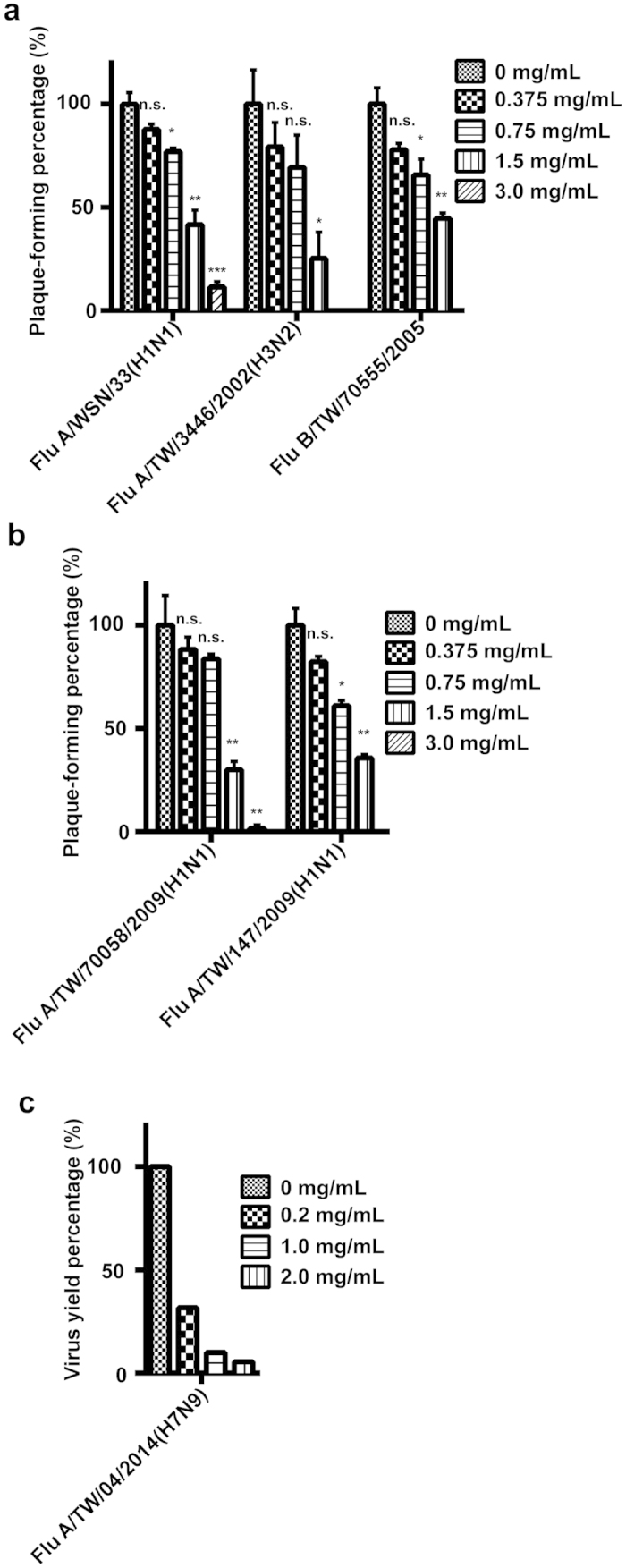
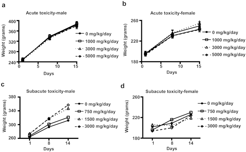
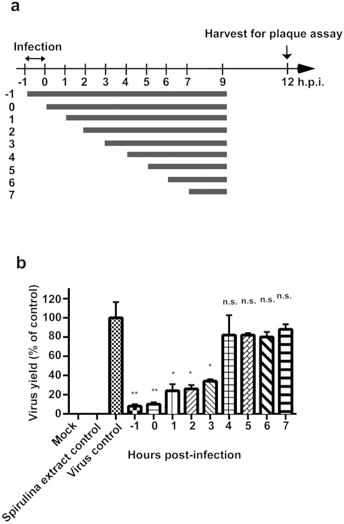
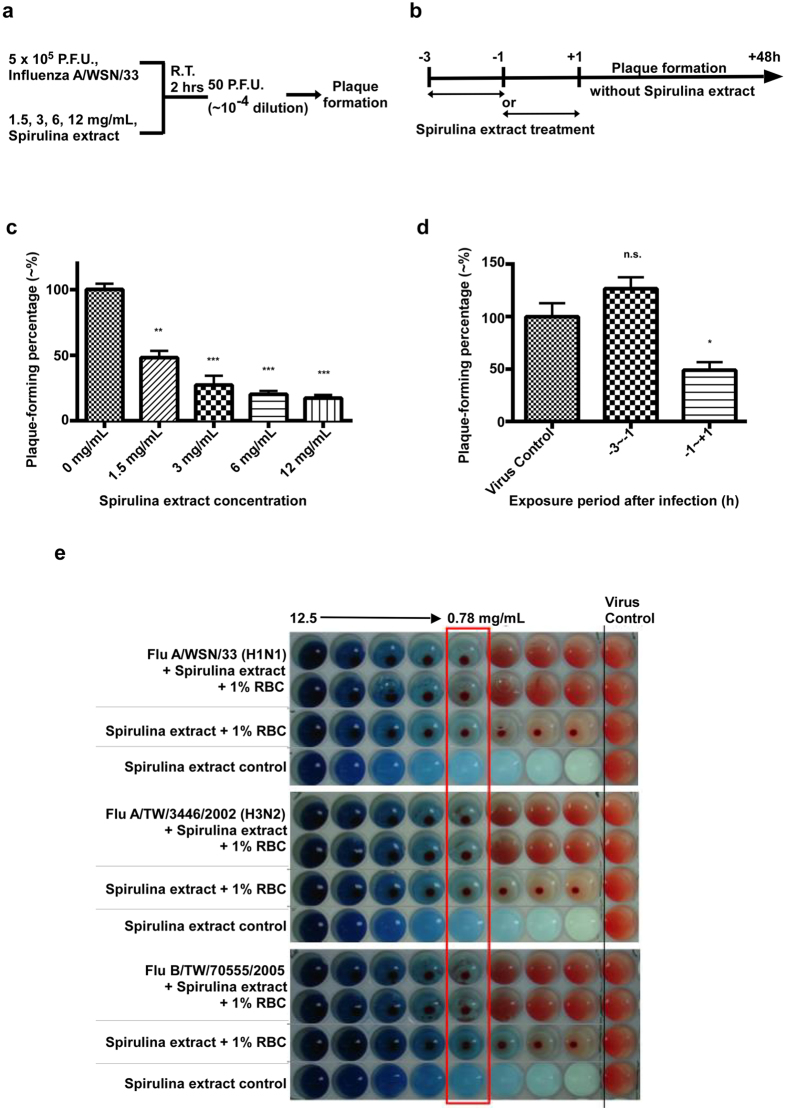
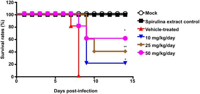
Influenza is one of the most common human respiratory diseases, and represents a serious public health concern. However, the high mutability of influenza viruses has hampered vaccine development, and resistant strains to existing anti-viral drugs have also emerged. Novel anti-influenza therapies are urgently needed, and in this study, we describe the anti-viral properties of a Spirulina (Arthrospira platensis) cold water extract. Anti-viral effects have previously been reported for extracts and specific substances derived from Spirulina, and here we show that this Spirulina cold water extract has low cellular toxicity, and is well-tolerated in animal models at one dose as high as 5,000 mg/kg, or 3,000 mg/kg/day for 14 successive days. Anti-flu efficacy studies revealed that the Spirulina extract inhibited viral plaque formation in a broad range of influenza viruses, including oseltamivir-resistant strains. Spirulina extract was found to act at an early stage of infection to reduce virus yields in cells and improve survival in influenza-infected mice, with inhibition of influenza hemagglutination identified as one of the mechanisms involved. Together, these results suggest that the cold water extract of Spirulina might serve as a safe and effective therapeutic agent to manage influenza outbreaks, and further clinical investigation may be warranted.


Figures

Figure 1
Figure 1. The cold water extract of Spirulina (Arthrospira platensis) inhibits viral replication and plaque formation in a broad range of influenza strains in vitro.
(a) The influenza virus strains A/WSN/33(H1N1), A/TW/3446/02(H3N2), and B/TW/70555/05, and (b) the oseltamivir-resistant strains A/TW/70058/09(H1N1) and A/TW/147/09(H1N1), propagated on MDCK cells, all exhibited dose-dependent reductions in virus plaque formation after treatment with 0.375, 0.75, 1.5, and 3.0 mg/ml of Spirulina extract. Data represent the mean ± SD for three independent experiments. Statistical significance was determined by two-tailed unpaired t-test, and used as the basis to label results as ns, not significant; *P ≤ 0.05; **P ≤ 0.01; or ***P ≤ 0.001. (c) The TCID50 assay showed that Spirulina extract reduced virus production of the influenza A/TW/04/2014 (H7N9) virus.
Figure 2
Figure 2. Short-term acute and subacute oral toxicity for the Spirulina extract.
(a) 14-day acute oral toxicity study was conducted in Sprague-Dawley rats given 0, 1,000, 3,000, or 5,000 mg/kg of Spirulina extract. Body weight changes were tracked for (a) male and (b) female rats during the study period, and no significant changes were observed. (a) 14-day subacute study, in which Sprague-Dawley rats were subjected to repeated dosing with 750, 1,500, or 3,000 mg/kg/day of Spirulina extract for 14 consecutive days, was also conducted. Body weight changes for (c) male and (d) female rats were measured on Day 1, Day 8, and Day 14. All data are presented as mean ± SD (N = 10).
Figure 3
Figure 3. Spirulina extract acts to disrupt viral replication at an early stage of infection.
(a) A time of addition assay was conducted, in which 2.5 mg/mL of Spirulina extract was added at the indicated timepoints to MDCK cells infected with the A/WSN/33(H1N1) influenza strain. E0 medium was overlaid on cultures at 9 hours post-infection, and supernatants were harvested at 12 hours post-infection. (b) Virus yields were determined by the plaque assay, and reported as a percentage of untreated controls. Data represent the mean ± SD for two independent experiments. Statistical analysis was performed with two-tailed unpaired t-test, and the statistical significance was determined to be *P ≤ 0.05, **P ≤ 0.01, or ***P ≤ 0.001 for each treated group versus the virus control.
Figure 4
Figure 4. Spirulina extract inhibits influenza virus infection by disrupting hemagglutination.
(a) 5 × 105 PFUs of the influenza A/WSN/33(H1N1) virus was incubated with 1.5, 3, 6, or 12 mg/mL of Spirulina extract at room temperature for 2 hours, after which cultures underwent a 104-fold dilution to derive samples containing ~50 PFU and negligible amounts of Spirulina extract, which were then used in a plaque assay to assess viral viability. (b) Plaque assays showed that plaque formation percentages for Spirulina extract-treated viruses were significantly lower than untreated controls. Statistical analysis was performed with the two-tailed unpaired t-test, and used as the basis to label results as ns, not significant; *P ≤ 0.05; **P < 0.01; ***P < 0.001. (c) Monolayer MDCK cells in 6-well plates were either treated with 3 mg/mL of Spirulina extract for 2 hours, after which the culture medium was replaced with fresh medium containing no Spirulina extract (the −3 ~ −1 group), or treated with 3 mg/mL of Spirulina extract just prior to and during viral adsorption (the −1 ~ +1 group). 50 P.F.U. of the influenza A/WSN/33(H1N1) virus were added to cultures at Hour 0, and a plaque assay was conducted to assess viral viability after 48 hours of incubation. (d) Plaque assays showed that the plaque formation percentage of the −3 ~ −1 group was comparable to the untreated virus control, while plaque formation was significantly inhibited in the −1 ~ +1 group. Statistical analysis was performed with the two-tailed unpaired t-test, and used as the basis to label results as ns, not significant; *P ≤ 0.05; **P ≤ 0.01; or ***P ≤ 0.001 (e) Varying concentrations of Spirulina extract were added to 96-well plates containing 1% of guinea pig RBCs and 4 HA units of influenza A/WSN/33(H1N1), A/TW/3446/02(H3N2), or B/TW/70555/05 viruses. In the virus control column at far right, no Spirulina extract was added. Results showed that concentrations of Spirulina extract above 0.78 mg/mL were capable of inhibiting influenza hemagglutination.
Figure 5
Figure 5. Spirulina extract improves survival rates in influenza-infected mice.
Six-week-old female BALB/c mice were inoculated with 2 × 104 PFUs of the influenza A/WSN/33(H1N1) virus, after receiving Spirulina extract 4 hours prior to infection by oral gavage. Spirulina extract was again administered at 6 hours post-infection and twice daily thereafter for 4 days, at respective daily doses of 10, 25, or 50 mg/kg. Survival rates were improved in mice treated with Spirulina extract, as compared to vehicle-treated controls that did not receive Spirulina extract. Statistical analyses of the survival curves were performed using the Log-rank (Mantel-Cox) test compared each Spirulina extract treated group to the vehicle-treated group (n = 5 mice per group). *P < 0.05, **P < 0.01.

References

    1. Taubenberger J. K. & Morens D. M. The pathology of influenza virus infections. Annu. Rev. Pathol. 3, 499–522 (2008). - PMC - PubMed
    1. Tamerius J. et al. Global influenza seasonality: reconciling patterns across temperate and tropical regions. Environ. Health Perspect. 119, 439–445 (2011). - PMC - PubMed
    1. Johnson N. P. & Mueller J. Updating the accounts: global mortality of the 1918–1920 “Spanish” influenza pandemic. Bull. Hist. Med. 76, 105–115 (2002). - PubMed
    1. Dawood F. S. et al. Estimated global mortality associated with the first 12 months of 2009 pandemic influenza A H1N1 virus circulation: a modelling study. Lancet Infect. Dis. 12, 687–695 (2012). - PubMed
    1. Tong S. et al. New world bats harbor diverse influenza A viruses. PLoS pathogens 9, e1003657, 10.1371/journal.ppat.1003657 (2013). - DOI - PMC - PubMed

Publication types

Substances