

Monocyte-derived macrophages exhibit distinct and more restricted HIV-1 integration site repertoire than CD4(+) T cells
- PMID: 27067385
- PMCID: PMC4828718
- DOI: 10.1038/srep24157
Monocyte-derived macrophages exhibit distinct and more restricted HIV-1 integration site repertoire than CD4(+) T cells
Abstract
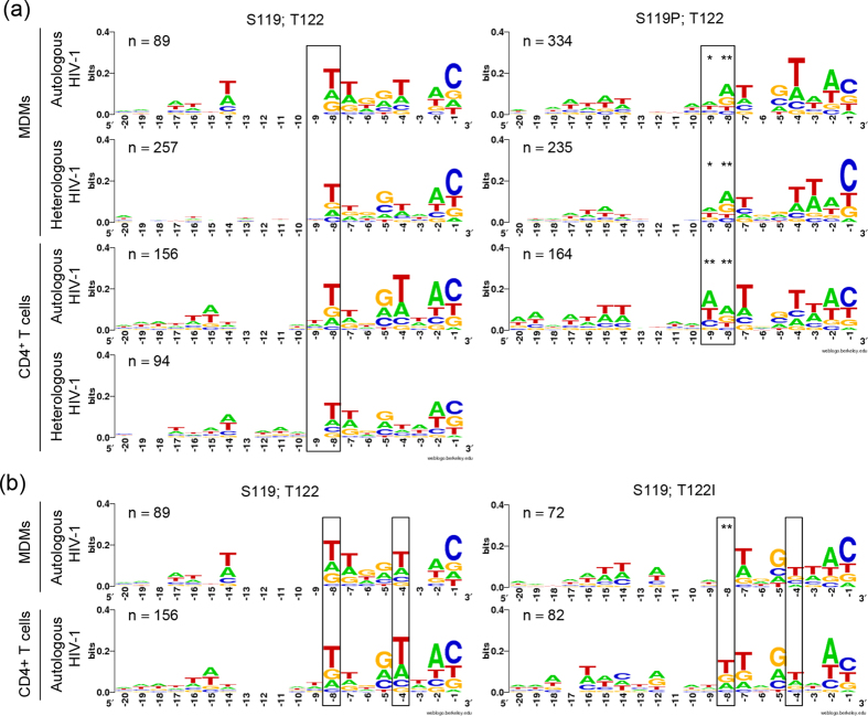
The host genetic landscape surrounding integrated HIV-1 has an impact on the fate of the provirus. Studies analysing HIV-1 integration sites in macrophages are scarce. We studied HIV-1 integration site patterns in monocyte-derived macrophages (MDMs) and activated CD4(+) T cells derived from seven antiretroviral therapy (ART)-treated HIV-1-infected individuals whose cells were infected ex vivo with autologous HIV-1 isolated during the acute phase of infection. A total of 1,484 unique HIV-1 integration sites were analysed. Their distribution in the human genome and genetic features, and the effects of HIV-1 integrase polymorphisms on the nucleotide selection specificity at these sites were indistinguishable between the two cell types, and among HIV-1 isolates. However, the repertoires of HIV-1-hosting gene clusters overlapped to a higher extent in MDMs than in CD4(+) T cells. The frequencies of HIV-1 integration events in genes encoding HIV-1-interacting proteins were also different between the two cell types. Lastly, HIV-1-hosting genes linked to clonal expansion of latently HIV-1-infected CD4(+) T cells were over-represented in gene hotspots identified in CD4(+) T cells but not in those identified in MDMs. Taken together, the repertoire of genes targeted by HIV-1 in MDMs is distinct from and more restricted than that of CD4(+) T cells.
Figures


Similar articles
-
Different kinetics of viral replication and DNA integration in the main HIV-1 cellular reservoirs in the presence and absence of integrase inhibitors.Antiviral Res. 2018 Dec;160:165-174. doi: 10.1016/j.antiviral.2018.10.017. Epub 2018 Oct 26. Antiviral Res. 2018. PMID: 30420339
-
Quantitative PCR used to assess HIV-1 integration and 2-LTR circle formation in human macrophages, peripheral blood lymphocytes and a CD4+ cell line.Virol J. 2010 Dec 3;7:354. doi: 10.1186/1743-422X-7-354. Virol J. 2010. PMID: 21129188 Free PMC article.
-
Characterization of the single cycle replication of HIV-1 expressing Gaussia luciferase in human PBMCs, macrophages, and in CD4(+) T cell-grafted nude mouse.J Virol Methods. 2016 Feb;228:95-102. doi: 10.1016/j.jviromet.2015.11.019. Epub 2015 Nov 27. J Virol Methods. 2016. PMID: 26640121
-
Experimental approaches to the study of HIV-1 latency.Nat Rev Microbiol. 2007 Feb;5(2):95-106. doi: 10.1038/nrmicro1580. Nat Rev Microbiol. 2007. PMID: 17224919 Review.
-
HIV-1 regulation of latency in the monocyte-macrophage lineage and in CD4+ T lymphocytes.J Leukoc Biol. 2010 Apr;87(4):575-88. doi: 10.1189/jlb.0409264. Epub 2009 Oct 2. J Leukoc Biol. 2010. PMID: 19801499 Review.
Cited by
-
Impact of chromatin on HIV-1 latency: a multi-dimensional perspective.Epigenetics Chromatin. 2025 Mar 8;18(1):9. doi: 10.1186/s13072-025-00573-x. Epigenetics Chromatin. 2025. PMID: 40055755 Free PMC article. Review.
-
Spontaneous reactivation of latent HIV-1 promoters is linked to the cell cycle as revealed by a genetic-insulators-containing dual-fluorescence HIV-1-based vector.Sci Rep. 2018 Jul 5;8(1):10204. doi: 10.1038/s41598-018-28161-y. Sci Rep. 2018. PMID: 29977044 Free PMC article.
-
HIV infection results in clonal expansions containing integrations within pathogenesis-related biological pathways.JCI Insight. 2018 Jul 12;3(13):e99127. doi: 10.1172/jci.insight.99127. JCI Insight. 2018. PMID: 29997284 Free PMC article.
-
Development of a Novel In Vitro Primary Human Monocyte-Derived Macrophage Model To Study Reactivation of HIV-1 Transcription.J Virol. 2021 Sep 9;95(19):e0022721. doi: 10.1128/JVI.00227-21. Epub 2021 Sep 9. J Virol. 2021. PMID: 34287050 Free PMC article.
-
HIV-1 integration sites in CD4+ T cells during primary, chronic, and late presentation of HIV-1 infection.JCI Insight. 2021 May 10;6(9):e143940. doi: 10.1172/jci.insight.143940. JCI Insight. 2021. PMID: 33784259 Free PMC article.
References
-
- Dalgleish A. G. et al.. The CD4 (T4) antigen is an essential component of the receptor for the AIDS retrovirus. Nature 312, 763–767 (1984). - PubMed
-
- Klatzmann D. et al.. T-lymphocyte T4 molecule behaves as the receptor for human retrovirus LAV. Nature 312, 767–768 (1984). - PubMed
-
- Koenig S. et al.. Detection of AIDS virus in macrophages in brain tissue from AIDS patients with encephalopathy. Science (New York, N.Y.) 233, 1089–1093 (1986). - PubMed
-
- Maddon P. J. et al.. The T4 gene encodes the AIDS virus receptor and is expressed in the immune system and the brain. Cell 47, 333–348 (1986). - PubMed
-
- Alkhatib G. et al.. CC CKR5: a RANTES, MIP-1alpha, MIP-1beta receptor as a fusion cofactor for macrophage-tropic HIV-1. Science (New York, N.Y.) 272, 1955–1958 (1996). - PubMed
Publication types
MeSH terms
LinkOut - more resources
Full Text Sources
Other Literature Sources
Research Materials
