. 2016 Apr;140 Suppl 1(Suppl 1):S27-36.
doi: 10.1016/S0049-3848(16)30095-0.

Activated tumor cell integrin αvβ3 cooperates with platelets to promote extravasation and metastasis from the blood stream

Affiliations

Activated tumor cell integrin αvβ3 cooperates with platelets to promote extravasation and metastasis from the blood stream

Martin R Weber et al. Thromb Res. 2016 Apr.

Abstract

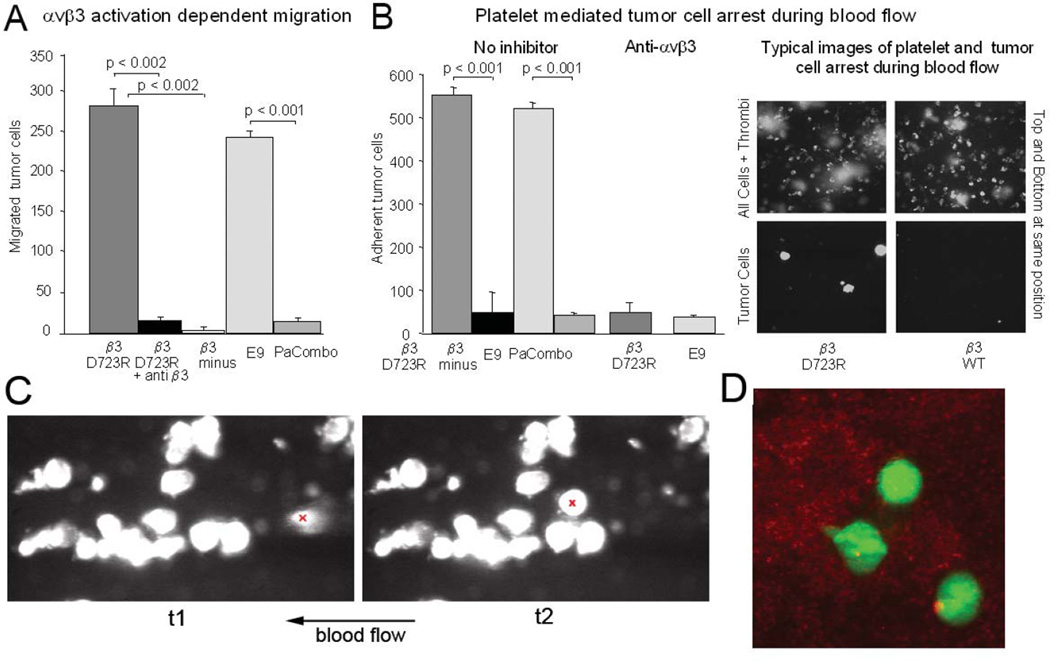
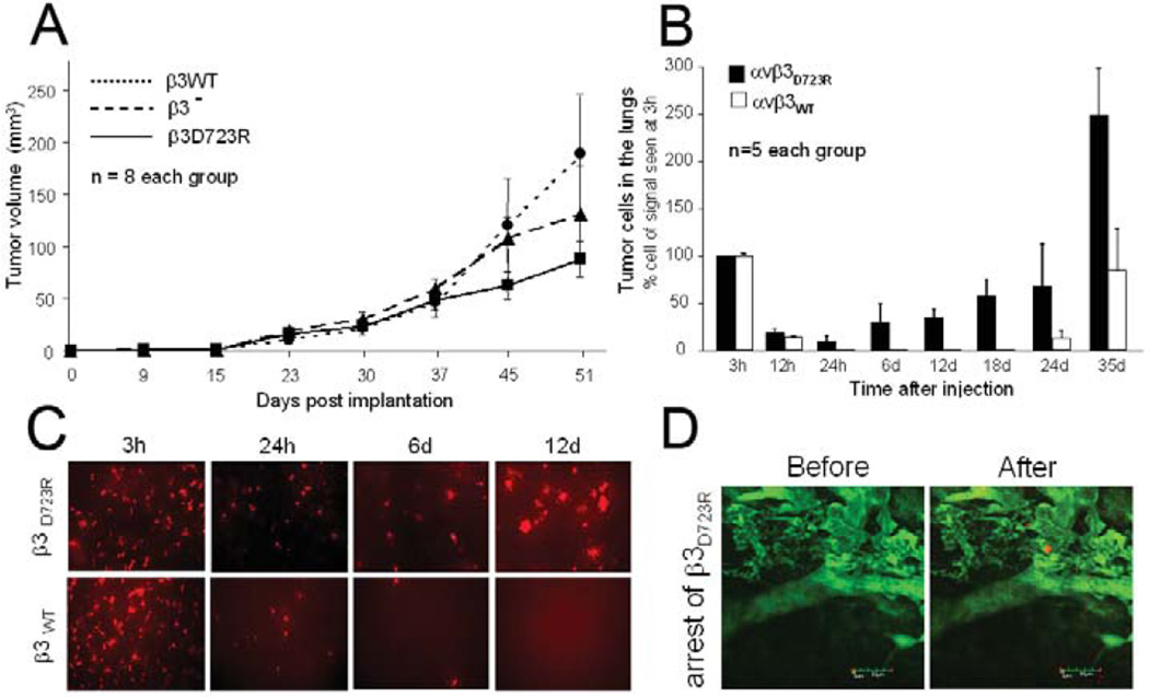
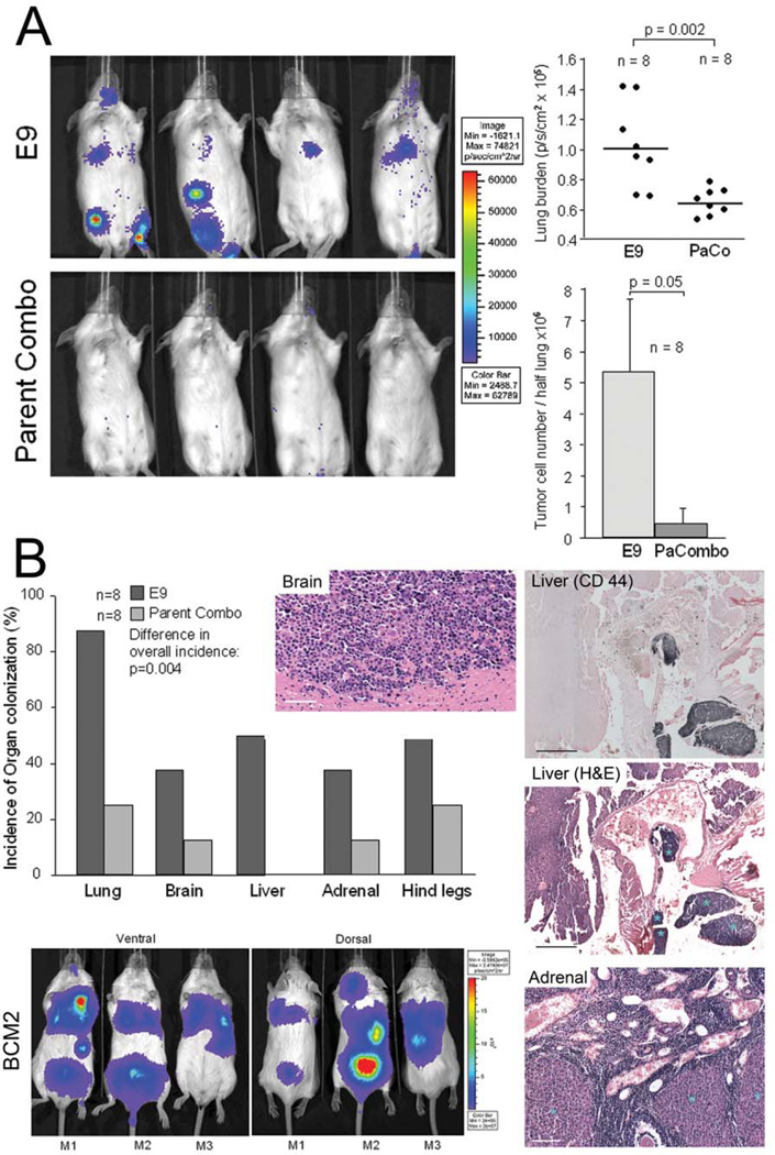
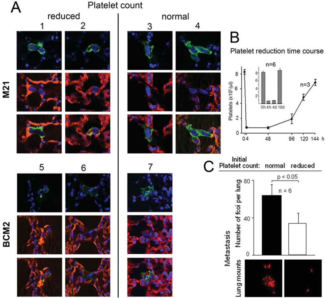
Metastasis is the main cause of death in cancer patients, and understanding mechanisms that control tumor cell dissemination may lead to improved therapy. Tumor cell adhesion receptors contribute to cancer spreading. We noted earlier that tumor cells can expressing the adhesion receptor integrin αvβ3 in distinct states of activation, and found that cells which metastasize from the blood stream express it in a constitutively high affinity form. Here, we analyzed steps of the metastatic cascade in vivo and asked, when and how the affinity state of integrin αvβ3 confers a critical advantage to cancer spreading. Following tumor cells by real time PCR, non-invasive bioluminescence imaging, intravital microscopy and histology allowed us to identify tumor cell extravasation from the blood stream as a rate-limiting step supported by high affinity αvβ3. Successful transendothelial migration depended on cooperation between tumor cells and platelets involving the high affinity tumor cell integrin and release of platelet granules. Thus, this study identifies the high affinity conformer of integrin αvβ3 and its interaction with platelets as critical for early steps during hematogenous metastasis and target for prevention of metastatic disease.

Keywords: Blood stream; Extravasation; Integrin activation; Metastasis; Platelets.


Figures

Figure 1
Figure 1. Expression of activated integrin αvβ3 in subpopulations of heterogeneous breast cancer cells and their interaction with platelets during blood flow
A, Haptotactic migration toward immobilized fibrinogen requires activated integrin αvβ3 in MDA-MB 435 cells. Cells selected for lack of β3 expression (β3 minus) failed to migrate in contrast to their counterparts reconstituted with the constitutively activating β3mutant D723R (p<0.002). Migration of β3 mutant D723R expressing cells is strongly inhibited by blocking anti-β3 antibody (mab M21-3 at 50 µg/ml) (p<0.002). E9, a random clone of MDA-MB 435 cells expresses high affinity αvβ3, while Parent Combo, a combination of 20 MDA-MB-435 clones has low affinity αvβ3 (p<0.001) (16 hr migration at 37C). Ooverall expression levels of the integrin are similar across all 435 variants (Fig S1). B, Tumor cell attachment to collagen I during blood flow relies on high-affinity integrin αvβ3 (blocked by anti αvβ3 mab VNR1-27.1) and involves platelets. The right panel shows images at predefined positions within a flow chamber, of MDA-MB 435 β3D723R or β3WT expressing cells during blood flow (top images: green fluorescence of arrested leukocytes and platelet aggregates; bottom images: red fluorescence of arrested tumor cells (dsRed) at the same positions). Arrested tumor cells are quantified at 50 predefined positions during ongoing perfusion, after the same number of tumor cells for each cell type were allowed to pass through the chamber. C, Dymanic tumor cell-platelet interaction during blood flow. Conditions as in B, but using M21 human melanoma cells in mouse blood. Shown are two consecutive frames of a video microcopy analysis (link to video to be inserted here). The flow field shows a group of tumor cells already attached in a platelet dependent manner. An additional tumor cell, entering the view field, arrests on an existing platelet-tumor heteroaggregate. This cell is marked with a red ‘x’ in the frames in C, at time points 1 and 2 (t1, t2). Tumor cells can anchor on attached platelets, and sometimes carry platelets at their surface which facilitate tumor cell attachment during blood flow. D, Dual-color confocal analysis of tumor cell-platelet interaction during blood flow. M21 melanoma cells (green) interacting with attached, thrombus-forming platelets (red). Upon contact with platelets, tumor cells extend pseudopods (cell in the middle). Statistical significance in A,B was assessed by paired, one-tailed t-tests.
Figure 2
Figure 2. Activation of tumor cell integrin αvβ3 promotes initial steps of target organ colonization from the blood stream but is not required for primary tumor growth
A, Tumor growth in the mammary fat pad of MDA-MB 435 cells lacking β3 expression (β3), or reconstituted with β3 wild type (β3WT) versus constitutively activating mutant β3 (β3D723R). Tumor volumes in SCID mice (n=8/group) were measured with calipers and calculated as (a2×b)/2. B, Quantification of tumor cells in SCID mouse lungs at varying time points after tail vein injection of 1×10⁵ dsRed tagged MDA-MB 435 cells expressing activated αvβ3D723R or non-activated αvβ3WT. Tumor cells were quantified by real time PCR of human alu sequences within the lung tissue. Initial signal at 3h post injection was nearly identical for both cell types and reflects tumor cells within the pulmonary microcirculation, most of which are cleared during the first day. Bars indicate numbers of lung associated tumor cells (n=5 mice) shown as % (± SEM) of the initial tumor cell number at 3 hours post injection. Cell numbers are based on PCR standard curves, obtained from lung homogenates spiked with known tumor cell numbers. C, Lung whole mounts 3 to 12 hrs after tail vein injection of dsRed tagged tumor cells as in B, visualizing the tumor cells by fluorescence microscopy (10×). Representative fields are shown. D, Arrest of β3D723R expressing cells within the pulmonary microvasculature observed by intravital microscopy. Lung allografts were grown in dorsal skin fold chambers on SCID mice, allowed to vascularize for 14 days before superfusion with TNFα (50µl at 1µg/ml) 2.5 hours before i.v. injection of 2.5×10⁶ dsRed tagged tumor cells together with FITC-dextran (500 kDa) to visualize the vasculature. Circulation and adhesive events were recorded by intravital video microscopy (Leitz Biomed). Bar, 50 µm.
Figure 3
Figure 3. Integrin αvβ3 activation promotes hematogenous metastasis to multiple target organs
A, Left: Metastatic activity of MDA-MB 435 clone E9 expressing intrinsically activated integrin αvβ3 compared to 20-clone pool Parent Combo expressing non-activated αvβ3. Non-invasive bioluminescence imaging of SCID mice 14 days after tail vein injection of 1×10⁵ F-luc tagged tumor cells. Right: Quantification of metastatic burden in the lung region by non-invasive bioluminescence imaging, 14 days after injecting 1×10⁵ tumor cells (Top), or by real time PCR 34 days after injecting 1×10⁶ tumor cells to challenge the system and following the tumor cells by human alu sequences (Bottom). Each data point in the top panel represents signal from one mouse (n=8), the vertical line indicates median signal in each group. Bars in the lower panel show average values (+/− STDEV) for each group (n=8). Clone E9 caused significantly higher metastatic lung burden than clone pool Parent Combo (p=0.002 by bioluminescence imaging 14 days after 1×10⁵ injected tumor cells; p= 0.05 by alu PCR 34 days after 1×10⁶ injected tumor cells). Statistical significance was assessed by paired, one-tailed t-tests. B, Comparison of target organ colonization by tumor cells expressing activated versus non-activated αvβ3. Incidence of tumor cell burden in the lungs, brain, liver, adrenal glands and bone (hind legs), 56 days after tail vein injection of 1×10⁵ clone E9 cells versus clone pool Parent Combo. Organ associated metastatic burden was detected in excised organs by ex vivo bioluminescence imaging, n= 8 mice / group. The presence of metastatic lesions was verified by histology (right panels). Brain lesion H&E staining, Bar 80 µm. Liver lesion, top panel identifies tumor lesions by immunohistochemistry with anti human CD44 (Mab 29.7) (dark blue), bottom panel shows a neighboring section stained with H&E and lesions marked with blue asterisks, Bar 200 µm. Adrenal gland section shown by H&E staining, lesions marked with light blue asterisks, Bar 150 µm. Mouse images: Multiple target organ colonization validated with polyclonal cell population BCM2 which express activated integrin αvβ3. Non-invasive bioluminescence imaging of metastasis development in distinct target organs, 56 days after tail vein injection of 2.5×10⁵ tumor cells. Three representative mice are shown (ventral and dorsal). Metastatic burden in the lungs, liver, brain, spine, and adrenal gland was confirmed by bioluminescence imaging of the excised organs, and by histology as above.
Figure 4
Figure 4. Activated integrin αvβ3 promotes tumor cell transendothelial migration in a platelet dependent manner
A, Platelets and high affinity tumor cell integrin αvβ3 together promote tumor cell transendothelial migration in vitro. In the presence of platelets, MDA-MB 435 cell variants expressing high affinity αvβ3D723R, or intrinsically activated αvβ3 (BCM2), penetrated monolayers of human lung microvascular endothelial cells more efficiently than tumor cells with low affinity αvβ3WT (p=0.03). Platelet activation with TRAP-6 (thrombin receptor activating peptide-6 and PAR1 agonist) before incubation with tumor cells did not further enhance the stimulatory effect. TRAP-6 alone without platelets, or exhausted platelets (stimulated with TRAP-6, allowed to release their α granules, and washed before addition to tumor cells), did not enhance tumor cell transendothelial migration. B, Platelet releaseate reduces endothelial monolayer integrity in the presence of tumor cells. Intact human pulmonary endothelial layers (initial transendothelial resistance >30Ω/cm2) were incubated with BCM2 cells, and washed human platelets stimulated with ADP, or their ATP induced releasates were added. Transendothelial resistance was measured at the indicated time points. Insert shows damaged endothelial layer with a tumor cell crossing over into a denuded area (yellow arrow).
Figure 5
Figure 5. Platelets promote tumor cell extravasation and metastasis from the blood stream in vivo
A, Tumor cell extravasation from the pulmonary microvasculature in SCID mice with reduced or normal platelet counts. Thrombocytopenia was induced with anti-murine platelet GP1bα 4 hrs before i.v. injection of M21 melanoma or BMC2 breast cancer cells. Extravasation was analyzed by confocal microscopy on day 3, revealing tumor cells (human CD44 in green) inside the vasculature (CD31, PECAM in red) in thrombocytopenic mice (mouse# 1,2,5,6), or outside the vasculature in mice with normal platelet counts (mouse# 3,4,7). B, Platelet reduction time course. Thrombocytopenia was induced as in A (n=3), and platelet counts measured at the indicated time points. Insert shows platelet counts in mice in which experimental metastasis, measured in C (n=6). C, Thrombocytopenia reduces metastasis from the blood stream. SCID mice were injected with anti platelet glycoprotein GPIbα as above, or PBS as control (n=6). Four hours later, 1×10⁵ sdRed tagged BCM2 tumor cells were injected into the tail vein and lung metastases counted microscopically in whole mounts of each lung lobe, 18 days later. Bars denote the number of metastatic foci per lung ± SEM, counted regardless of size. Images below show dsRed tumor cell lesions in lung whole mounts representative for each experimental group. Mice with normal platelet counts had significantly more metastases (p<0.05) than thrombocytopenic mice. Generally, lesions in control mice were also considerably larger than lesions in anti-platelet treated mice. Differences in metastatic burden were confirmed by real time PCR of human alu sequences within the lung tissue (not shown). Statistical significance was assessed by paired, one-tailed t-tests.

Similar articles

Cited by

References

    1. Hynes RO. The emergence of integrins: a personal and historical perspective. Matrix Biol. 2004;23:333–340. - PMC - PubMed
    1. Arnaout MA, Goodman SL, Xiong JP. Structure and mechanics of integrin-based cell adhesion. Curr Opin Cell Biol. 2007;19:495–507. - PMC - PubMed
    1. Ginsberg MH, Partridge A, Shattil SJ. Integrin regulation. Curr Opin Cell Biol. 2005;17:509–516. - PubMed
    1. Luo BH, Carman CV, Springer TA. Structural Basis of Integrin Regulation and Signaling. Annu Rev Immunol. 2007;25:619–647. - PMC - PubMed
    1. Felding-Habermann B. Integrin adhesion receptors in tumor metastasis. Clin Exp Metastasis. 2003;20:203–213. - PubMed

Publication types

Substances