. 2016 May 24;84(6):1806-1814.
doi: 10.1128/IAI.00130-16. Print 2016 Jun.

The Metalloprotease Mpl Supports Listeria monocytogenes Dissemination through Resolution of Membrane Protrusions into Vacuoles

Affiliations

The Metalloprotease Mpl Supports Listeria monocytogenes Dissemination through Resolution of Membrane Protrusions into Vacuoles

Diego E Alvarez et al. Infect Immun. .

Abstract

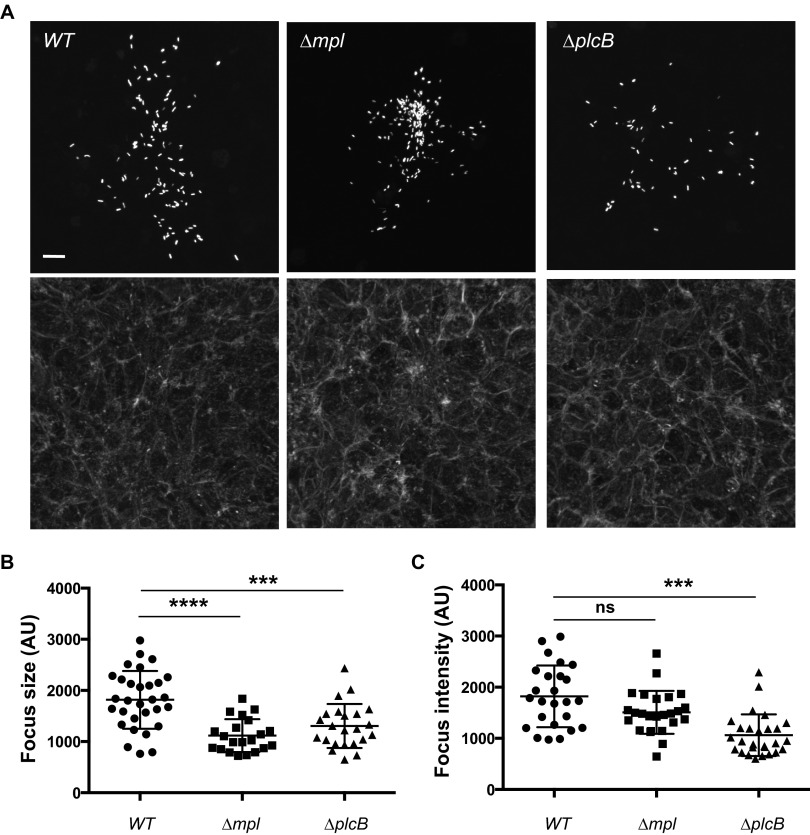
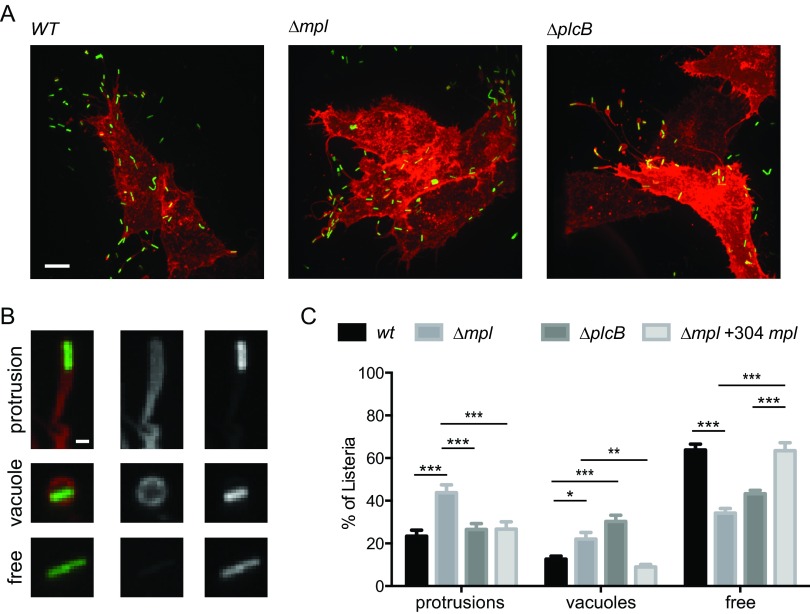
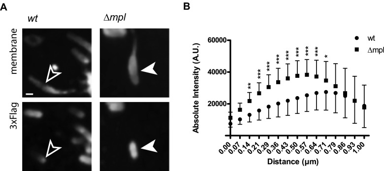
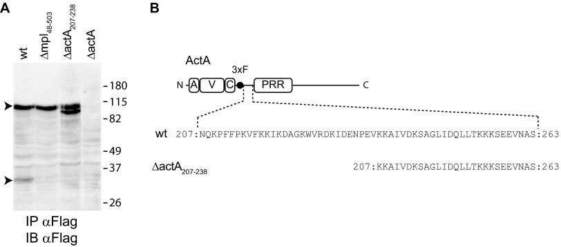
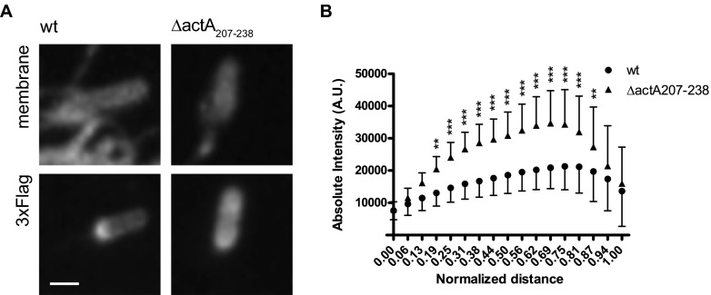
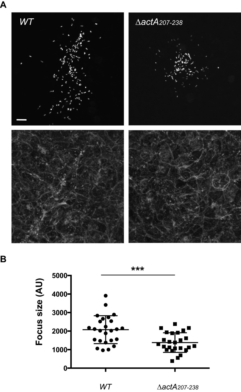
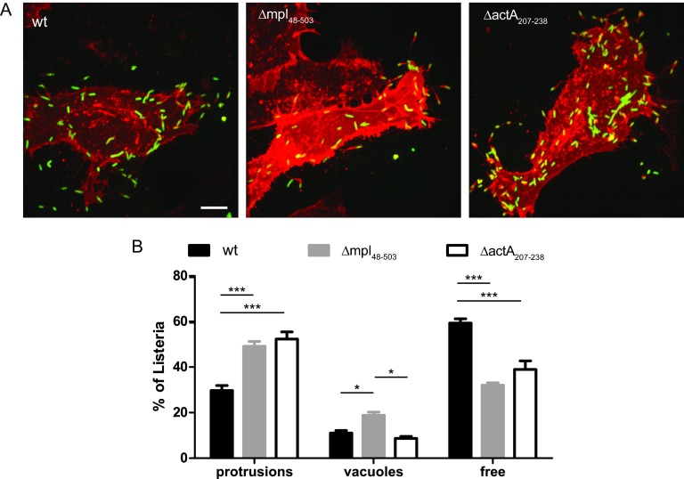
Listeria monocytogenes is an intracellular pathogen that disseminates within the intestinal epithelium through acquisition of actin-based motility and formation of plasma membrane protrusions that project into adjacent cells. The resolution of membrane protrusions into vacuoles from which the pathogen escapes results in bacterial spread from cell to cell. This dissemination process relies on the mlp-actA-plcB operon, which encodes ActA, a bacterial nucleation-promoting factor that mediates actin-based motility, and PlcB, a phospholipase that mediates vacuole escape. Here we investigated the role of the metalloprotease Mpl in the dissemination process. In agreement with previous findings showing that Mpl is required for PlcB activation, infection of epithelial cells with the ΔplcB or Δmpl strains resulted in the formation of small infection foci. As expected, the ΔplcB strain displayed a strong defect in vacuole escape. However, the Δmpl strain showed an unexpected defect in the resolution of protrusions into vacuoles, in addition to the expected but mild defect in vacuole escape. The Δmpl strain displayed increased levels of ActA on the bacterial surface in protrusions. We mapped an Mpl-dependent processing site in ActA between amino acid residues 207 to 238. Similar to the Δmpl strain, the ΔactA207-238 strain displayed increased levels of ActA on the bacterial surface in protrusions. Although the ΔactA207-238 strain displayed wild-type actin-based motility, it formed small infection foci and failed to resolve protrusions into vacuoles. We propose that, in addition to its role in PlcB processing and vacuole escape, the metalloprotease Mpl is required for ActA processing and protrusion resolution.


Figures

FIG 1
FIG 1
Characterization of the infection foci formed in cells infected with the wild type (WT) or the mpl or plcB mutant. (A) Representative images showing monolayers of confluent HeLa 229 cells infected with GFP-expressing L. monocytogenes for 6 h (top, GFP-expressing bacteria; bottom, phalloidin staining). Bar, 10 μm. (B) Sizes of the infection foci (AU, arbitrary units) as determined by computer-assisted analysis of images like those shown in panel A. One-way analysis of variance (ANOVA) with Tukey's posttest: Δmpl versus WT strain, P < 0.0001 (****); ΔplcB versus WT strain, P < 0.001 (***). (C) Average GFP intensity per infection focus as determined by computer-assisted analysis of images shown in panel A. One-way ANOVA with Tukey's posttest: Δmpl versus WT strain, not significant (ns); ΔplcB versus WT strain, P < 0.001 (***).
FIG 2
FIG 2
Protrusion resolution and vacuole escape in cells infected with the mpl and plcB mutants. (A) Representative images showing HeLa 229 cells transiently transfected with the plasma membrane-targeted dsRed constructs and infected with GFP-expressing L. monocytogenes for 6 h (green, bacteria; red, membrane). Bar, 10 μm. (B) Representative images of spreading bacteria in protrusions (top panel), in double-membrane vacuoles (middle panel), and not associated with membrane marker (bottom panel). Left, merged image (green, bacteria; red, plasma membrane). Middle, plasma membrane only. Right, bacteria. Bar, 1 μm. (C) Percentages of spreading bacteria in protrusions, in vacuoles, and free in adjacent cells in cells infected with wild-type bacteria or mpl or plcB mutants. Data are presented as means and standard deviations for three independent samples and were analyzed by two-way ANOVA with Tukey's posttest using PRISM 5 (GraphPad Software). ***, P < 0.001; **, P < 0.01; *, P < 0.05.
FIG 3
FIG 3
Distribution of ActA on the surface of wild-type and Δmpl mutant bacteria forming protrusions. (A) Representative images showing the distribution of 3×Flag-ActA on the surface of bacteria in protrusions. Top panels, plasma membrane. Bottom panels, FLAG staining. Bar, 1 μm. (B) Quantitative analysis of ActA distribution on the bacterial surface. The absolute intensity of the fluorescence signal in the FLAG channel is represented against the normalized distance from the front pole. Data are represented as means and standard deviations and were analyzed by two-way ANOVA using PRISM 5 (GraphPad Software). ***, P < 0.001; **, P < 0.01; *, P < 0.05.
FIG 4
FIG 4
Mapping of an Mpl-dependent processing site in ActA. (A) Western blot of ActA 3×F immunoprecipitated (IP) from cell lysates infected with L. monocytogenes wild-type, Δmpl48–503, ΔactA207–238, or ΔactA strains. Arrowheads indicate ActA peptides of 90 and 30 kDa. Apparent molecular weights (in thousands) in the protein ladder are indicated on the right. IB, immunoblot. (B) Schematic representation of 3×Flag-ActA and in-frame deletion of ActA residues 207 to 238. Numbers indicate the first and last residues.
FIG 5
FIG 5
Distribution of ActA on the surface of wild-type and ΔactA207–238 mutant bacteria forming protrusions. (A) Representative images showing the distribution of 3×Flag-ActA on the surface of bacteria in protrusions. Top panels, plasma membrane. Bottom panels, FLAG staining. Bar, 1 μm. (B) Quantification of ActA distribution on the bacterial surface. The absolute intensity of the FLAG signal is represented against the normalized distance from the front pole. Data are represented as means and standard deviations and were analyzed by two-way ANOVA using PRISM 5 (GraphPad Software). ***, P < 0.001; **, P < 0.01; *, P < 0.05.
FIG 6
FIG 6
Characterization of the infection foci formed in cells infected with the wild-type or ΔactA207–238 mutant strains. (A) Representative images showing monolayers of confluent HeLa 229 cells infected with GFP-expressing L. monocytogenes for 6 h (top, GFP-expressing bacteria; bottom, phalloidin staining). Bar, 10 μm. (B) Sizes of the infection foci as determined by computer-assisted analysis of images like those shown in panel A. Unpaired t test: ΔactA207–238 versus WT strain, P < 0.001 (***).
FIG 7
FIG 7
Protrusion resolution and vacuole escape in cells infected with the mpl and ΔactA207–238 mutant strains. (A) Representative images showing protrusion and vacuole formation in HeLa 229 cells transiently transfected with the plasma membrane-targeted dsRed constructs and infected with GFP-expressing L. monocytogenes for 6 h (green, bacteria; red, membrane). Bar, 10 μm. (B) Percentages of spreading bacteria in protrusions, in vacuoles, and free in adjacent cells in cells infected with the wild-type bacteria or with mpl or ΔactA207–238 mutant strains. Data are presented as means and standard deviations for three independent samples and were analyzed by two-way ANOVA with Tukey's posttest using PRISM 5 (GraphPad Software). ***, P < 0.001; *, P < 0.05.

References

    1. Gouin E, Welch MD, Cossart P. 2005. Actin-based motility of intracellular pathogens. Curr Opin Microbiol 8:35–45. doi:10.1016/j.mib.2004.12.013. - DOI - PubMed
    1. Robbins JR, Barth AI, Marquis H, de Hostos EL, Nelson WJ, Theriot JA. 1999. Listeria monocytogenes exploits normal host cell processes to spread from cell to cell. J Cell Biol 146:1333–1350. doi:10.1083/jcb.146.6.1333. - DOI - PMC - PubMed
    1. Tilney LG, Portnoy DA. 1989. Actin filaments and the growth, movement, and spread of the intracellular bacterial parasite, Listeria monocytogenes. J Cell Biol 109:1597–1608. doi:10.1083/jcb.109.4.1597. - DOI - PMC - PubMed
    1. Dragoi AM, Agaisse H. 2015. The class II phosphatidylinositol 3-phosphate kinase PIK3C2A promotes Shigella flexneri dissemination through formation of vacuole-like protrusions. Infect Immun 83:1695–1704. doi:10.1128/IAI.03138-14. - DOI - PMC - PubMed
    1. Kuehl CJ, Dragoi AM, Talman A, Agaisse H. 2015. Bacterial spread from cell to cell: beyond actin-based motility. Trends Microbiol 23:558–566. doi:10.1016/j.tim.2015.04.010. - DOI - PMC - PubMed

Publication types

MeSH terms

Substances

LinkOut - more resources