. 2016 Apr 12:6:24452.
doi: 10.1038/srep24452.

Dynamic interaction of SARAF with STIM1 and Orai1 to modulate store-operated calcium entry

Affiliations

Dynamic interaction of SARAF with STIM1 and Orai1 to modulate store-operated calcium entry

Letizia Albarran et al. Sci Rep. .

Abstract

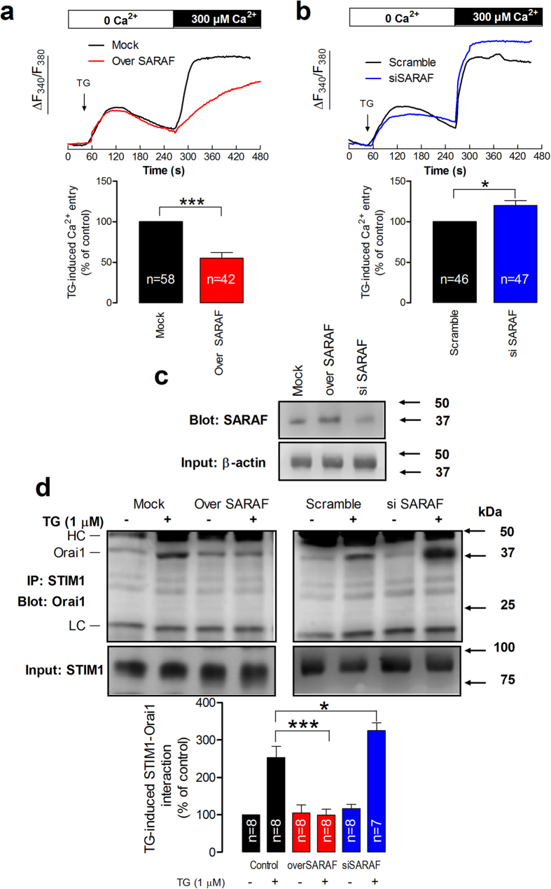
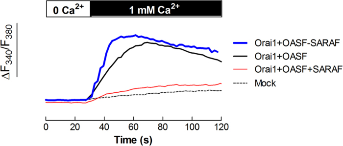
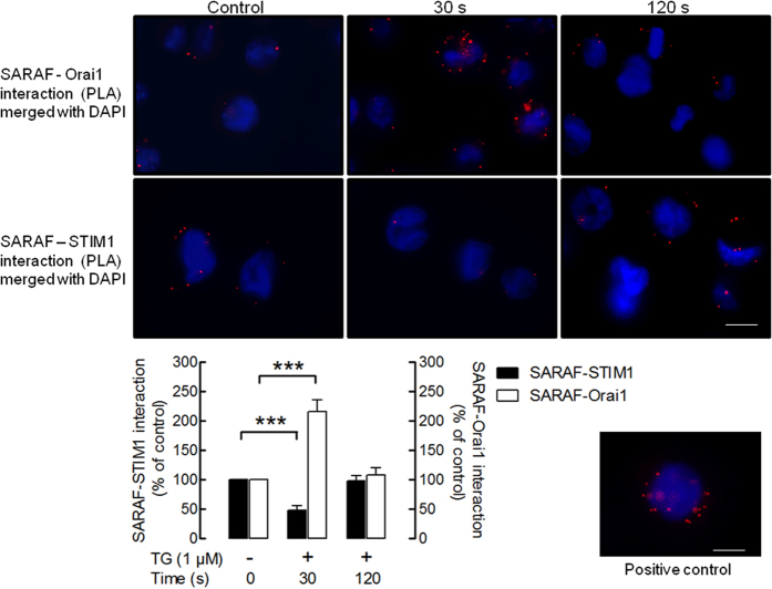
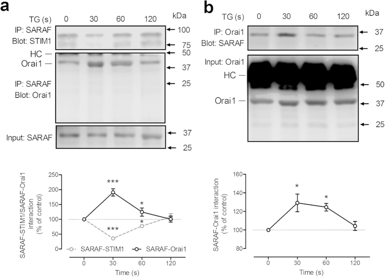
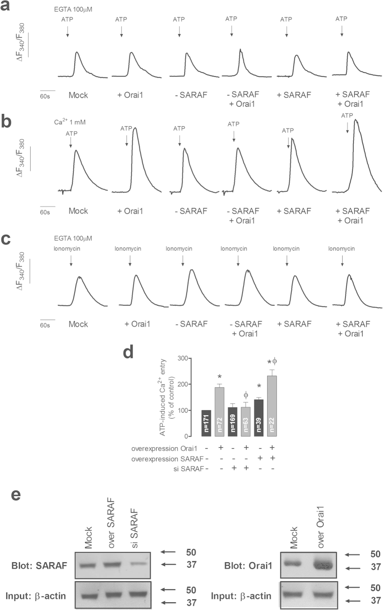
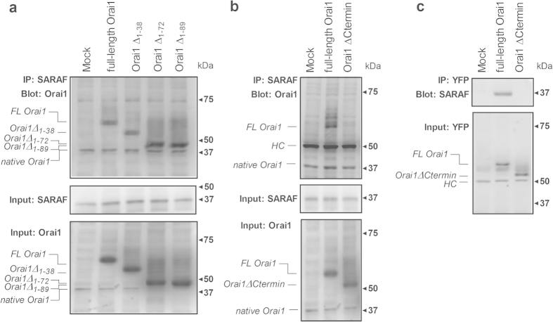
Ca(2+) influx by store-operated Ca(2+) channels is a major mechanism for intracellular Ca(2+) homeostasis and cellular function. Here we present evidence for the dynamic interaction between the SOCE-associated regulatory factor (SARAF), STIM1 and Orai1. SARAF overexpression attenuated SOCE and the STIM1-Orai1 interaction in cells endogenously expressing STIM1 and Orai1 while RNAi-mediated SARAF silencing induced opposite effects. SARAF impaired the association between Orai1 and the Orai1-activating small fragment of STIM1 co-expressed in the STIM1-deficient NG115-401L cells. Cell treatment with thapsigargin or physiological agonists results in direct association of SARAF with Orai1. STIM1-independent interaction of SARAF with Orai1 leads to activation of this channel. In cells endogenously expressing STIM1 and Orai1, Ca(2+) store depletion leads to dissociation of SARAF with STIM1 approximately 30s after treatment with thapsigargin, which paralleled the increase in SARAF-Orai1 interaction, followed by reinteraction with STIM1 and dissociation from Orai1. Co-expression of SARAF and either Orai1 or various N-terminal deletion Orai1 mutants did not alter SARAF-Orai1 interaction; however, expression of C-terminal deletion Orai1 mutants or blockade of the C-terminus of Orai1 impair the interaction with SARAF. These observations suggest that SARAF exerts an initial positive role in the activation of SOCE followed by the facilitation of SCDI of Orai1.


Figures

Figure 1
Figure 1. SARAF regulates TG-induced SOCE and STIM1-Orai1 interaction.
(a,b) MEG-01 cells overexpressing SARAF and mock-treated cells (a) or MEG-01 cells transfected with si SARAF or scramble plasmid (b) were perfused with a Ca2+-free medium (100 μM EGTA added) and then stimulated with TG (1 μM) followed by reintroduction of external Ca2+ (final concentration 300 μM) to initiate Ca2+ entry. Data are original traces representative of 42-58 experiments. Values are expressed as described in methods. The bar graph represents TG-induced Ca2+ entry as mean ± SEM and data are presented as percentage of control. (c) Cells overexpressing SARAF, treated with siSARAF and mock-treated cells were lysed and subjected to Western blotting with anti-SARAF antibody, followed by reprobing with anti-actin antibody for protein loading control. (d) MEG-01 cells overexpressing SARAF or transfected with si SARAF and their respective controls were stimulated with TG (1 μM) in a Ca2+-free medium (100 μM EGTA added) and three min later lysed. Whole cell lysates were immunoprecipitated (IP) with anti-STIM1 antibody and immunoprecipitates were subjected to 10% SDS-PAGE and subsequent Western blotting with a specific anti-Orai1 (aa 288–301 (Sigma)) antibody. Membranes were reprobed with the antibody used for immunoprecipitation for protein loading control. The panels show results from one experiment representative of 6–7 others. Molecular masses indicated on the right were determined using molecular-mass markers run in the same gel. HC, heavy chain of the antibody used for immunoprecipitation. The bar graph represents the quantification of STIM1-Orai1 association in resting and TG-treated cells. Results are recorded as arbitrary optical density units, expressed as mean ± S.E.M. and presented as percentage of control. * and *** represent p < 0.05 and p < 0.001, as compared to their respective controls.
Figure 2
Figure 2. SARAF impairs OASF-dependent Orai1 function.
NG115-401L cells were transfected with pEYPF-Orai1 and pEYPF-OASF in the absence of presence of SARAF or siSARAF or were mock treated, as indicated. After 48h cells were perfused with a Ca2+-free medium (100 μM EGTA added) followed by reintroduction of external Ca2+ (final concentration 1 mM) to initiate constitutive Ca2+ entry. Data are original traces representative of 42–58 experiments. Values are expressed as described in methods.
Figure 3
Figure 3. Dynamic interaction of SARAF with Orai1 and STIM1.
MEG-01 cells were suspended in HBS containing 300 μM CaCl2 and were left untreated or stimulated with TG (1 μM) for 30 or 120 s. The cells were then fixed with 4% ice-cold paraformaldehyde and blocked using blocking solution from the Duolink In Situ Red kit® from Sigma. Samples were incubated with primary antibodies: rabbit anti-SARAF and mouse anti-STIM1 or mouse anti-Orai1 (SAB4200273, Sigma) antibodies. A Duolink assay was subsequently performed according to the manufacturer’s instructions as described in Material and Methods. After performing the PLA procedure, images were taken with an Eclipse TE300 fluorescence microscope. Red spots represent individual dimers. The positive control is shown in the lower right corner. The bars indicate 10 μm. The bar graphs indicate association between SARAF and STIM1 or Orai1 expressed as the mean ± S.E.M. of 100–200 cells. The quantification of PLA signals was performed using ImageJ software. ***p < 0.001.
Figure 4
Figure 4. Time-course of TG-induced SARAF-STIM1 and SARAF-Orai1 co-immunoprecipitation.
MEG-01 cells (a) and NG115-401L cells (b) were stimulated with TG (1 μM) in the presence of 300 μM extracellular Ca2+. Samples were removed 30s before the addition of TG and 30, 60 and 120 s after the treatment with TG. Whole cell lysates were immunoprecipitated (IP) with the indicated antibody and immunoprecipitates were subjected to 10% SDS-PAGE and subsequent Western blotting with specific anti-Orai1 (aa 288–301 (Sigma)), anti-SARAF and anti-STIM1 antibodies. Membranes were reprobed with the antibody used for immunoprecipitation for protein loading control. The panels show results from one experiment representative of 5 others. Molecular masses indicated on the right were determined using molecular-mass markers run in the same gel. HC, heavy chain of the antibody used for immunoprecipitation. Data represent the quantification of SARAF-Orai1 and SARAF-STIM1 association in resting and TG-treated cells. Results are recorded as arbitrary optical density units, expressed as mean ± S.E.M. and presented as percentage of control (resting cells). * and *** represent p < 0.05 and p < 0.001, as compared to controls.
Figure 5
Figure 5. SARAF regulates Orai1 channel function.
(a–c) NG115-401L cells were transfected with Orai1, SARAF, siSARAF or empty vector as control (Mock). Cells were perfused with a Ca2+-free medium (100 μM EGTA added) (a,c) or a medium containing 1 mM Ca2+ (b) and then stimulated with ATP (100 μM; (a,b)) or ionomycin (2 μM, (c)). Data are original traces representative of 22–171 experiments. (d) The bar graph represents ATP-evoked Ca2+ entry estimated as the integral of the rise in [Ca2+]c above basal for 1½ min after the addition of ATP in the presence of external Ca2+, corrected by subtraction of the integral over the same period for stimulation with ATP in the absence of external Ca2+. Data are expressed as mean ± SEM and presented as percentage of control (mock-treated cells). * and ϕ represent p < 0.05 as compared to ATP-induced Ca2+ entry in mock-treated controls or cells overexpressing Orai1, respectively. (e) Immunoblot analysis of SARAF and Orai1 expressed in NG115-401L cells before or after treatment with siSARAF or expression plasmids for SARAF and Orai1 using anti-SARAF antibody and anti-Orai1 antibody (aa 288–301 (Sigma)). Membranes were reprobed with anti-β-actin antibody for protein loading control. The panel shows results from one experiment representative of 3 others. Molecular masses indicated on the right were determined using molecular-mass markers run in the same gel.
Figure 6
Figure 6. Analysis of the Orai1 site that interacts with SARAF.
NG115-401L cells were transfected with SARAF alone or in combination with either pEYFP-full-length Orai1 (FL-Orai1), the N-terminal deletion mutants (pEYFP-Orai1 ΔN1–38 (Orai1 ΔN1–38), ΔN1–72 (Orai1 ΔN1–72) and ΔN1–89 (Orai1 ΔN1–89) (a), pEYFP-Orai1 C-terminal deletion mutant (Orai1Ctermin, amino acids 1–260 (b,c) or empty vector (Mock), as indicated. After 48 h cells were lysed and the whole cell lysates were immunoprecipitated (IP) with anti-SARAF antibody (a,b) or anti-YFP antibody (c). Immunoprecipitates were subjected to 10% SDS-PAGE and subsequent Western blotting with a specific anti-Orai1 (amino acids 288–301, Sigma) antibody (a), anti-Orai1 (ab177021, Abcam) antibody (b) or anti-SARAF antibody (c). Membranes were reprobed with the antibody used for immunoprecipitation for protein loading control (middle panels). (a,b) Alternatively, the cell lysates were subjected to 10% SDS-PAGE and subsequent Western blotting with anti-Orai1 antibody (specific for amino acids 288–301; panel a, bottom) or with anti-Orai1 (ab177021, Abcam; panel b, bottom). HC, heavy chain of the antibody used for immunoprecipitation. The panels show results from one experiment representative of 3 others. Molecular masses indicated on the right were determined using molecular-mass markers run in the same gel.

Similar articles

Cited by

References

    1. Roos J. et al. STIM1, an essential and conserved component of store-operated Ca2+ channel function. J Cell Biol 169, 435–445 (2005). - PMC - PubMed
    1. Liou J. et al. STIM is a Ca2+ sensor essential for Ca2+-store-depletion-triggered Ca2+ influx. Curr Biol 15, 1235–1241 (2005). - PMC - PubMed
    1. Feske S. et al. A mutation in Orai1 causes immune deficiency by abrogating CRAC channel function. Nature 441, 179–185 (2006). - PubMed
    1. Vig M. et al. CRACM1 multimers form the ion-selective pore of the CRAC channel. Curr Biol 16, 2073–2079 (2006). - PMC - PubMed
    1. Zhang S. L. et al. Genome-wide RNAi screen of Ca2+ influx identifies genes that regulate Ca2+ release-activated Ca2+ channel activity. Proc Natl Acad Sci USA 103, 9357–9362 (2006). - PMC - PubMed

Publication types

MeSH terms