. 2016 Apr 12:6:23978.
doi: 10.1038/srep23978.

ExtraPEG: A Polyethylene Glycol-Based Method for Enrichment of Extracellular Vesicles

Affiliations

ExtraPEG: A Polyethylene Glycol-Based Method for Enrichment of Extracellular Vesicles

Mark A Rider et al. Sci Rep. .

Abstract

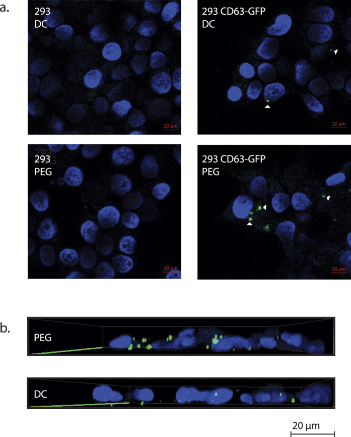
Initially thought to be a means for cells to eliminate waste, secreted extracellular vesicles, known as exosomes, are now understood to mediate numerous healthy and pathological processes. Though abundant in biological fluids, purifying exosomes has been challenging because their biophysical properties overlap with other secreted cell products. Easy-to-use commercial kits for harvesting exosomes are now widely used, but the relative low-purity and high-cost of the preparations restricts their utility. Here we describe a method for purifying exosomes and other extracellular vesicles by adapting methods for isolating viruses using polyethylene glycol. This technique, called ExtraPEG, enriches exosomes from large volumes of media rapidly and inexpensively using low-speed centrifugation, followed by a single small-volume ultracentrifugation purification step. Total protein and RNA harvested from vesicles is sufficient in quantity and quality for proteomics and sequencing analyses, demonstrating the utility of this method for biomarker discovery and diagnostics. Additionally, confocal microscopy studies suggest that the biological activity of vesicles is not impaired. The ExtraPEG method can be easily adapted to enrich for different vesicle populations, or as an efficient precursor to subsequent purification techniques, providing a means to harvest exosomes from many different biological fluids and for a wide variety of purposes.


Figures

Figure 1
Figure 1. Polyethylene glycol enriches vesicles containing exosome proteins.
(a) Nanoparticle tracking analysis identified particles in all PEG treatments. The greatest number of particles were recovered using 12% PEG (dashed line represents baseline particle count: particles harvested from conditioned medium assessed by NTA prior to PEG treatment; bounded by one standard deviation). (b) No differences in particle size between groups were detected. (c) Purity was plotted as a ratio of particles recovered to micrograms of protein in sample. Sample purity peaked when PEG concentrations of 6 to 8% were used. (d,e) Lysates were evaluated for exosome protein. Equal-volume (35uL) or equal-mass (8 μg) gel-loading was used to evaluate exosome recovery and purity, respectively. (f,g) Blots were Ponceau stained to evaluate total protein, and compared to (h) fetal bovine serum protein (FBS) alone (protein recovered from PEG-treated, unconditioned culture medium, with 10% FBS). All approximate protein masses are represented in kilodaltons (kDa).
Figure 2
Figure 2. PEG-based vesicle enrichment is comparable or superior to standard harvest methods.
(a) All harvest methods recovered more particles than differential centrifugation (DC). (b) Particle sizes were consistent with the reported size distributions of exosomes. (c) The average sample purity of the 8% PEG + wash group was not statistically different from the DC group (p > 0.05), but was purer than those of the commercial products (p < 0.01). (d) Of the PEG-based methods, 8% PEG + wash and 12% PEG + wash produced the strongest evidence of exosomes by western blot. (e) Western analyses showed that purity of 8% PEG + wash group was comparable to DC, and superior to commercial methods. (f,g) Ponceau staining demonstrated that no method removed all serum protein contaminants, however, the 8% PEG + wash method greatly reduced the most prominent contaminants relative to other treatments. *A non-specific >50 kDa band appeared in the ExoQuick sample lane (d). It was subsequently confirmed to not reflect the presence of TSG101 (see Fig. 3). TEI, Total Exosome Isolation (Life Technologies). All approximate protein masses are represented in kilodaltons (kDa).
Figure 3
Figure 3. Highly pure extracellular vesicles can be enriched using PEG.
(a) Western blot was used to assess the purity of samples from conditioned serum-free media (SFM). Abundances of exosome markers in PEG-treated samples were comparable to the sucrose cushion isolate. Probing for TSG101 produced a non-specific band at the 55–60 kDa marker; the non-specific band appeared consistently in samples containing only stock medium with 10% FBS, and in the bovine serum albumin (BSA) negative control. (b) Gels were coomassie-stained to compare total protein isolated from sucrose cushion and PEG methods. (c) The 8% PEG + wash method produced highly pure samples, comparable to the sucrose cushion isolates (no statistical difference, p = 0.169). (d) No differences in particle size were observed between treatment groups. (e) Presence of exosome-sized, cup-shaped vesicles was verified by electron microscopy. FBS, fetal bovine serum. DC, differential centrifugation. TEI, Total Exosome Isolation (Life Technologies). All approximate protein masses are represented in kilodaltons (kDa).
Figure 4
Figure 4. Extracellular vesicle proteins identified following ExtraPEG.
Mass spectrometry analysis of ExtaPEG-harvested protein from HeLa cells identified 519 unique proteins. Approximately 97% (499 of 517) of ExtraPEG-identified proteins were found in the Vesiclepedia database of described human extracellular vesicle proteins. Additionally, 95% (492 of 517) of the proteins were previously characterized as exosome proteins. Of the 519 mass spectrometry identifications, 517 were used for this analysis as there were two sets of proteins for which a single gene name was used by the Exocarta and Vesiclepedia databases to represent multiple protein accession numbers (i.e.: HLA-A represented 1A68_HUMAN and 1A69_HUMAN; HLA-B represented 1B15_HUMAN and 1B55_HUMAN).
Figure 5
Figure 5. Efficient uptake of ExtraPEG isolated exosomes.
(a) Confocal microscopy images of HEK293 cells. Cells were incubated for 24 hours with exosomes produced by HEK293 cells or HEK293 cells stably expressing CD63-GFP. Exosomes were harvested using ExtraPEG (PEG) or differential centrifugation (DC). (b) Three-dimensional image of combined z-stacks taken from cells exposed to PEG or DC-harvested CD63-GFP exosomes.
Figure 6
Figure 6
Extracellular vesicles harvested by ExtraPEG contain small RNAs. Exosome and cellular RNA were analyzed using the Agilent 2100 Bioanalyzer. (a) Gel-like image displaying RNA separated by size, for cell lysate (CL), differential centrifugation (DC), and the ExtraPEG (PEG) methods samples. (b) Electropherograms of RNA profiles from CL, DC, and PEG samples. [nt], nucleotide length. [FU], fluorescent units.

References

    1. Trams E. G., Lauter C. J., Salem N. Jr. & Heine U. Exfoliation of membrane ecto-enzymes in the form of micro-vesicles. Biochimica et biophysica acta 645, 63–70 (1981). - PubMed
    1. Colombo M., Raposo G. & Thery C. Biogenesis, secretion, and intercellular interactions of exosomes and other extracellular vesicles. Annual review of cell and developmental biology 30, 255–289, doi: 10.1146/annurev-cellbio-101512-122326 (2014). - DOI - PubMed
    1. Yanez-Mo M. et al. Biological properties of extracellular vesicles and their physiological functions. J Extracell Vesicles 4, 27066, doi: 10.3402/jev.v4.27066 (2015). - DOI - PMC - PubMed
    1. Meckes D. G. Jr., Menaker N. F. & Raab-Traub N. Epstein-Barr virus LMP1 modulates lipid raft microdomains and the vimentin cytoskeleton for signal transduction and transformation. Journal of virology 87, 1301–1311, doi: 10.1128/jvi.02519-12 (2013). - DOI - PMC - PubMed
    1. Meckes D. G. Jr. et al. Human tumor virus utilizes exosomes for intercellular communication. Proceedings of the National Academy of Sciences of the United States of America 107, 20370–20375, doi: 10.1073/pnas.1014194107 (2010). - DOI - PMC - PubMed

Publication types