. 2016 Sep 1;594(17):4753-68.
doi: 10.1113/JP272167. Epub 2016 Jul 3.

Quantifying sympathetic neuro-haemodynamic transduction at rest in humans: insights into sex, ageing and blood pressure control

Affiliations

Quantifying sympathetic neuro-haemodynamic transduction at rest in humans: insights into sex, ageing and blood pressure control

L J B Briant et al. J Physiol. .

Abstract

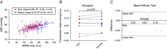
Key points: We have developed a simple analytical method for quantifying the transduction of sympathetic activity into vascular tone. This method demonstrates that as women age, the transfer of sympathetic nerve activity into vascular tone is increased, so that for a given level of sympathetic activity there is more vasoconstriction. In men, this measure decreases with age. Test-re-test analysis demonstrated that the new method is a reliable estimate of sympathetic transduction. We conclude that increased sympathetic vascular coupling contributes to the age-related increase in blood pressure that occurs in women only. This measure is a reliable estimate of sympathetic transduction in populations with high sympathetic nerve activity. Thus, it will provide information regarding whether treatment targeting the sympathetic nervous system, which interrupts the transfer of sympathetic nerve activity into vascular tone, will be effective in reducing blood pressure in hypertensive patients. This may provide insight into which populations will respond to certain types of anti-hypertensive medication.

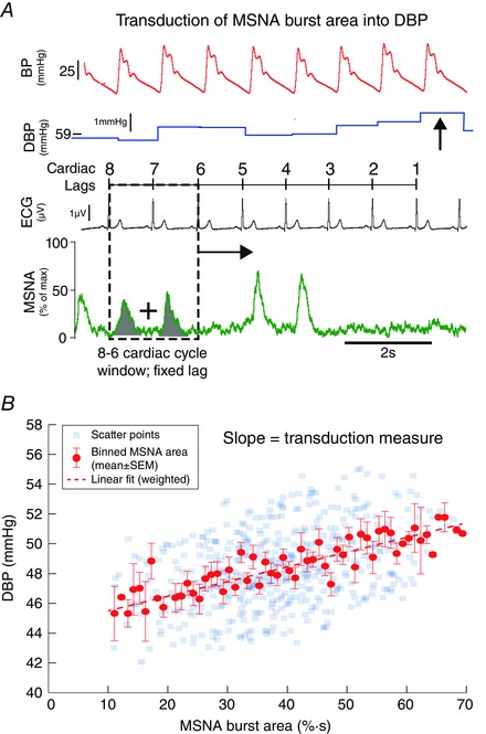
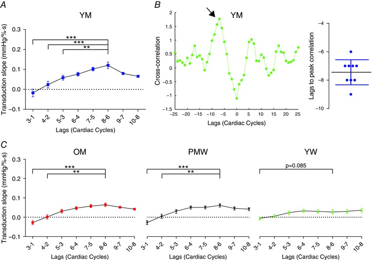
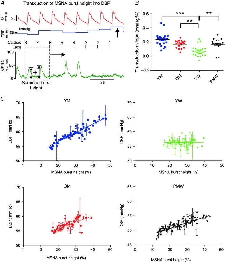
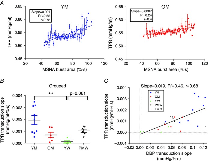
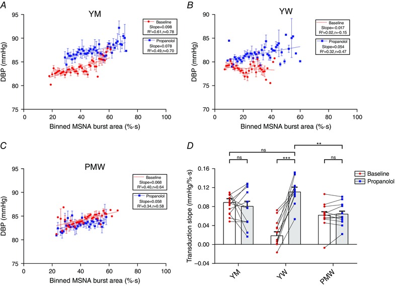
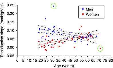
Abstract: Sex and age differences in the sympathetic control of resting blood pressure (BP) may be due to differences in the transduction of sympathetic nerve activity (SNA) into vascular tone. Current methods for dynamically quantifying transduction focus on the relationship between SNA and vasoconstriction during a pressor stimulus, which increases BP and may be contra-indicated in patients. We describe a simple analytical method for quantifying transduction under resting conditions. We performed linear regression analysis of binned muscle SNA burst areas against diastolic BP (DBP). We assessed whether the slope of this relationship reflects the transduction of SNA into DBP. To evaluate this, we investigated whether this measure captures differences in transduction in different populations. Specifically, we (1) quantified transduction in young men (YM), young women (YW), older men (OM) and postmenopausal women (PMW); and (2) measured changes in transduction during β-blockade using propranolol in YW, YM and PMW. YM had a greater transduction vs. OM (0.10 ± 0.01 mmHg (% s)(-1) , n = 23 vs. 0.06 ± 0.01 mmHg (% s)(-1) , n = 18; P = 0.003). Transduction was lowest in YW (0.02 ± 0.01 mmHg (% s)(-1) , n = 23) and increased during β-blockade (0.11 ± 0.01 mmHg (% s)(-1) ; P < 0.001). Transduction in PMW (0.07 ± 0.01 mmHg (% s)(-1) , n = 23) was greater compared to YW (P = 0.001), and was not altered during β-blockade (0.06 ± 0.01 mmHg (% s)(-1) ; P = 0.98). Importantly, transduction increased in women with age, but decreased in men. Transduction in women intersected that in men at 55 ± 1.5 years. This measure of transduction captures age- and sex-differences in the sympathetic regulation of DBP and may be valuable in quantifying transduction in disease. In particular, this measure may help target treatment strategies in specific hypertensive subpopulations.


Figures

Figure 1
Figure 1. Method of quantifying sympathetic neurovascular transduction into DBP in humans
A, for each DBP (arrowed), we summed MSNA burst area (shaded) in a 2 cardiac cycle window at a fixed lag (dashed window). In this example, the fixed lag is 8–6 cardiac cycles preceding the DBP. This ‘window’ was moved across the whole baseline file, associating each DBP with an MSNA burst area. B, these data were represented as a scatter plot for a fixed lag of 8–6 cardiac cycles. MSNA burst area was then binned into 1% s bins, and the corresponding DBP (mean ± SEM) plotted. A weighted linear regression was then fitted to these data, the slope of which gave our measurement of transduction (units of mmHg (% s)−1). MSNA, muscle sympathetic nerve activity; ECG, electrocardiogram; DBP, diastolic blood pressure; BP, blood pressure.
Figure 2
Figure 2. Determining optimal lag for analysis
To determine the cardiac cycle lag used for our analysis (see Fig. 1), we conducted the same analysis at different cardiac cycle lags, all of which yielded a ‘window’ of 2 cardiac cycles. A, for a sample population of 10 young men, the transduction was greatest at 8–6 cardiac lags. B, cross‐correlations of beat‐to‐beat MSNA burst area with diastolic blood pressure (DBP) in YM (n = 10). Example cross‐correlation in a participant; peak correlation (arrowed) occurred at a lag of −7 cardiac cycles. Grouped data revealed that correlation peaked at a lag of −7.44 ± 0.42 cardiac cycles. This supports our choice of lag as 8–6 cardiac cycles. C, we repeated the same lag analysis in OM, PMW and YW. For OM and PMW, 8–6 cardiac cycles produced peak transduction. In YW, transduction measured at 8–6 cardiac cycles was nearly significantly greater than at 3–1 (P = 0.082). Therefore, in all subsequent analysis in all groups, we used 8–6 cardiac cycles as our fixed choice of lag. YM, younger men; OM, older men; YW, younger women; PMW, postmenopausal women; one‐way repeated measures ANOVA, ** P < 0.01, *** P < 0.001.
Figure 3
Figure 3. Sympathetic neurovascular transduction of MSNA burst area into DBP in the sample population
Transduction of MSNA burst area into DBP was measured in all 83 participants. A, example transductions are depicted in a younger man (YM; a), younger woman (YW; b), older man (OM; c) and postmenopausal woman (PMW; d). B, grouped data for the transduction measurement revealed that transduction was reduced in YW compared to YM (P < 0.001), OM (P < 0.001) and PMW (P < 0.01). YM had the largest transduction measurement of all groups. There was no difference in the transduction measurement between OM and PMW (P = 0.32). C, R 2 of linear fits for each transduction slope. Goodness of fit for YW was low because of a lack of relationship between MSNA and DBP. ** P < 0.01, *** P < 0.001, ns = not significant (one‐way ANOVA; Kruskal–Wallis test with Dunn's multiple comparison).
Figure 4
Figure 4. Sympathetic neurovascular transduction of MSNA burst height into DBP in the sample population
Transduction of MSNA burst height into DPB was measured in all 83 participants. A, the method of calculating transduction of MSNA burst height into DBP. Each DBP was associated with summed MSNA burst heights over a fixed cardiac lag (8–6 cardiac cycles). Subsequent regression analysis was then conducted on these paired data points (as in Fig. 1 B). B, grouped data of transduction of MSNA burst height into DBP. C, example transduction regressions in a younger man (YM), younger woman (YW), older man (OM) and postmenopausal woman (PMW). ** P < 0.01, *** P < 0.001 (one‐way ANOVA; Kruskal–Wallis test with Dunn's multiple comparison).
Figure 5
Figure 5. Method of quantifying sympathetic neurovascular transduction into TPR
Transduction of MSNA burst area into total peripheral resistance (TPR) was also measured. Beat‐by‐beat TPR was calculated as MAP/stroke volume in a subpopulation of 9 YM, 6 OM, 5 YW and 6 PMW. For each beat‐by‐beat TPR, MSNA burst area was summed in a 2 cardiac cycle window at a lag of 8–6 cardiac cycles and associated with that TPR (similar to DBP in Fig. 1 A; see Methods). This window was moved across the whole baseline file, associating each TPR with an MSNA burst area. A, binned MSNA burst area (1% s bins) was plotted against the associated TPR (mean ± SEM). The transduction of MSNA burst area into TPR was taken as the slope of this relationship. Example for a younger male participant (left; with a steep slope/transduction) and an older male participant (right; with a gentler slope/transduction). B, grouped data for the transduction of MSNA burst area into TPR. C, transduction of MSNA burst area into TPR and DBP are linearly related. YM, younger men; OM, older men; YW, younger women; PMW, postmenopausal women; ** P < 0.01 (one‐way ANOVA; Kruskal–Wallis test with Dunn's multiple comparison).
Figure 6
Figure 6. Sympathetic neurovascular transduction during β‐blockade
Transduction of MSNA burst area into DBP was measured in a subset of 26 participants before (baseline) and after β‐blockade with propranolol. A–C, example regressions in a young man (YM), young woman (YW) and postmenopausal woman (PMW). D, grouped data revealed that propranolol did not change transduction in YM (P = 0.87; n = 12) or PMW (P = 0.98; n = 13) but increased transduction in YW (P < 0.001; n = 11). Following β‐blockade, YW had a greater transduction than both YM (P < 0.001) and PMW (P < 0.01). Two‐way repeated‐measures ANOVA, ** P < 0.01, *** P < 0.001, ns, not significant.
Figure 7
Figure 7. Test–re‐test analysis of transduction measurement
To determine the reproducibility of the measure of transduction of MSNA burst area into DBP we conducted a test–re‐test protocol on 8 participants. Transduction was measured during a baseline visit (test) and then re‐measured during a follow‐up visit on a different day (re‐test). A, example of test and re‐test transduction measurement for a participant. B, there was no difference between the test transduction and re‐test transduction (P = 0.25; paired t test). C, Bland–Altman test of agreement revealed that there was a small negative bias (which is lower than the mean differences in transduction between groups), and therefore good agreement between the variables.
Figure 8
Figure 8. Effects of sex and ageing on sympathetic neurovascular transduction
We plotted transduction of MSNA burst area into DBP against age for all 83 participants. In men, transduction increased with age and was well‐fitted by a linear regression (slope = −0.0011 mmHg (% s years)−1, R 2 = 0.31, Pearson r = −0.56; dashed lines). In contrast, transduction in women had a positive relationship with age (slope = 0.0014 mmHg (% s years)−1, R 2 = 0.46, Pearson r = 0.68). The slopes of these two regressions were statistically different (P < 0.001). Two outliers (circled) were removed from the analyses.

Comment in

Similar articles

Cited by

References

    1. Barnes JN, Hart EC, Curry TB, Nicholson WT, Eisenach JH, Wallin BG, Charkoudian N & Joyner MJ (2014). Aging enhances autonomic support of blood pressure in women. Hypertension 63, 303–308. - PMC - PubMed
    1. Bell C, Seals DR, Monroe MB, Day DS, Shapiro LF, Johnson DG & Jones PP (2001). Tonic sympathetic support of metabolic rate is attenuated with age, sedentary lifestyle, and female sex in healthy adults. J Clin Endocrinol Metab 86, 4440–4444. - PubMed
    1. Blessing WW (1997). The Lower Brainstem and Bodily Homeostasis, pp. 165–268. Oxford University Press, New York.
    1. Burnstock G (2008). Non‐synaptic transmission at autonomic neuroeffector junctions. Neurochem Int 52, 14–25. - PubMed
    1. Burnstock G (2009). Purinergic cotransmission. Exp Physiol 94, 20–24. - PubMed

Publication types